

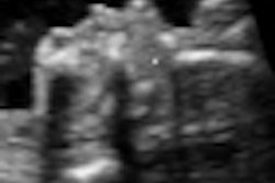
Incorporating gestational age (GA)-oriented morphology in biometry data could significantly increase the yield for ultrasound scanning of fetal malformations during the 6-16 weeks gestational age, according to research from the Ironfan-International Registry of Fetal Anomalies at Columbia University Medical Center in New York City.
"Time-oriented sonomorphology mapping is the most important component of the early malformation work-up. Fetal biometry has a minor impact," said Dr. Shraga Rottem of Columbia University at the 2007 American Institute of Ultrasound in Medicine (AIUM) meeting in New York City.
To investigate the potential effect of GA-oriented morphology mapping in addition to biometry in early malformation work-up, the Ironfan researchers began the study by utilizing personal experience, and calling for cases from 65 centers and practitioners worldwide of fetal anomalies, organs, and development charts detected during a 4-16 week sonogram.
They developed three comprehensive 4-16 weeks time-oriented databases, including a 4-16 weeks fetal anatomy and physiology database, a 6-16 weeks anomalies database, and a 4-16 weeks biometry database. As determined by ultrasound throughout pregnancy, fetal anomalies were classified into four classes:
- Type I: Early onset at constant gestational age
- Type II: Transient condition
- Type III: Variable onset or potentially unstable anomalies
- Type IV: Late onset anomalies
Using a specially designed time-oriented program, the researchers analyzed the 4-16 weeks database to determine the relative contributions of biometry and morphology-dysmorphology at the detection of a fetal anomaly at the earliest time of diagnosis between 6-16 weeks.
"We looked at the data and it was immediately obvious that morphology is what piqued the eye of the operator -- and not what came out from the US machine, which was the biometry," Rottem told AuntMinnie.com. "So here we have a situation where what comes from the routine scan from the machine has little contribution."
These conclusions were confirmed by a review of the literature, Rottem said.
"The literature confirmed our initial finding that the observation of morphology from the operator is much more important than what comes out from the data during the sonogram," he said.
Based on their own findings as well as literature review, the researchers determined that 161 organs, structures, and areas were detectable during a 4-16 weeks sonogram. In addition, 379 different types of fetal abnormalities may be detected at a 6-16 weeks sonogram, according to Rottem.
In 292 of the 379 (77%) types of fetal malformations studied, the 6-16 weeks malformation workup was based on morphology information alone, according to the researchers. Relying on biometry by itself was useful in only 87 anomalies (23%) of the cases.
The yield from combining morphology information and biometry is significantly increased for Type I anomalies, Rottem said. In addition, it was particularly useful in musculoskeletal and cardio/thorax malformations.
Rottem envisions a next generation of ob/gyn ultrasound systems that could move from malformation workup using fetal biometry charts to an approach that combines a early, time-oriented fetal sonomorphology system with results of normal or abnormal biometry.
This could be accomplished using Ironfan's database, linked to fetal biometry info and the ultrasound scanner, Rottem said.
 |
| The Ironfan Society's concept of incorporating a morphological system with biometry data to assist in time-oriented early fetal ultrasound scanning for malformations. Image courtesy of Dr. Shraga Rottem. |
A field test is currently being organized to link ultrasound machines with Ironfan's database, providing automatic feedback to the operator without user intervention, he said.
Ironfan would be interested in offering ultrasound vendors a link to the database for all its users, Rottem said.
"Our goal is to open this service via license to all the OEMs," he said. "This will allow the linking of their community of users and ultrasound systems to the database."
By Erik L. RidleyAuntMinnie.com staff writer
August 9, 2007
Related Reading
Fetal entertainment ultrasounds draw patient interest, March 20, 2007
Early prenatal ultrasound inadequate for detecting congenital heart malformations, June 30, 2006
Empty renal fossa on prenatal ultrasound indicates renal ectopia or agenesis, August 17, 2005
Fetal ear length may indicate chromosomal abnormality, April 11, 2002
Multinational study makes case for first-trimester sonography, June 12, 2001
Copyright © 2007 AuntMinnie.com








![Examples of ultrasound findings and techniques. (A) Images in a 39-year-old male patient with a mass in the left thigh. The mass is heterogeneous on the B-mode US image (compared with the patient in D) and showed increased microvascularity (superb microvascular imaging [SMI]) and shear-wave elastography (SWE) values. Undifferentiated pleomorphic sarcoma was diagnosed at biopsy (with pleomorphic rhabdomyosarcoma in surgical specimen). (B) Images in an 18-year-old male patient with a mass in the left leg. The mass is hypoechoic on the B-mode image, with no other findings suggestive of malignancy. The lesion is in contact with the cortex of the tibia, which is slightly irregular. CT revealed a doubtful anteromedial tibial erosion. The microvascular study demonstrated high vascularization, suggestive of malignancy. Periosteal Ewing sarcoma was diagnosed with both histologic and immunohistochemical confirmation. (C) Images in a 69-year-old female patient with a lump growing on the outside of the left leg. Multiple SWE examinations were performed (please note the high values obtained in the measurements, whereas the color map highlights the stiffness relative to adjacent tissues). SMI showed areas of increased vascularization to target for sampling. Undifferentiated spindle cell sarcoma was diagnosed at biopsy, with residual leiomyosarcoma in the surgical specimen after neoadjuvant therapy. (D) Images in a 56-year-old female patient with a mass in the right thigh. The mass is heterogeneous at both B-mode ultrasound (similar to patient A) and MRI (coronal T2-weighted spectral attenuated inversion recovery [SPAIR]; T1-weighted pre-contrast and postcontrast imaging), which even shows uptake after the administration of paramagnetic contrast material, which is traditionally suggestive of malignancy. Low values at SMI and elastography are suggestive of benignity. Spindle cell lipoma was diagnosed at biopsy, with atypical spindle cell lipomatous tumor in the surgical specimen.](https://img.auntminnie.com/mindful/smg/workspaces/default/uploads/2026/08/images-radiol250278fig2.APCFLSvX6p.jpg?auto=format%2Ccompress&dpr=2&fit=crop&h=167&q=70&w=250)


