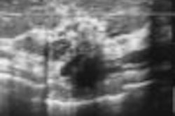
Real-time breast sonoelastography supports other imaging modalities in the evaluation of breast lesions, and shows promise for reducing biopsy rates, according to results from an Italian multicenter study.
"(Sonoelastography) complements conventional ultrasound and mammography in the evaluation of the breast lesion, mainly (in) BI-RADS 3 (patients)," said Dr. Giorgio Rizzatto of General Hospital in Gorizia, Italy. In BI-RADS 3 patients, "we have understood that you can move the follow-up schedule from six to 12 months, and also reduce the biopsy rate."
He presented the research during a scientific session at the 2007 RSNA meeting in Chicago.
To determine the clinical value of sonoelastography in the differential diagnosis of breast lesions in daily clinical practice, eight Italian institutions performed high-resolution ultrasound and sonoelastography using equipment from Hitachi Medical of Tokyo on a total of 784 women with a mean age of 52.5 years. The women had 874 lesions with a definitive diagnosis; 614 were benign, while 260 were malignant.
The ultrasound images were classified according to the BI-RADS criteria, while the sonoelastography images were assigned an elastographic score from 1 to 5 based on the distribution and degree of strain induced by light compression, according to Rizzatto.
Under the Italian classification system, a score of 1 indicated a three-layered pattern. A score of 2 indicated a lesion with even elastic pattern, while a score of 3 indicated a lesion with a mostly even elastic pattern, but with some areas of no strain. A score of 4 indicated that most of the lesion had no strain, while 5 represented a lesion with no strain.
Statistical analysis was performed by an independent institution. While the receiver operating curves (ROCs) showed a slightly better performance for ultrasound (area under the curve of 0.94 for BI-RADS) than for sonoelastography (0.90), the ROC curves demonstrated that elastography works better in lesions with a diameter of 15 mm or smaller, he said. The best results were obtained in lesions smaller than 5 mm in diameter.
The study team found that sonoelastography showed a very high specificity for benign lesions, including BI-RADS 3 lesions. When using the best cutoff point found between elasticity scores 3 and 4, the technique's negative predictive value was 98% for the whole series, 96.3% for all the BI-RADS 3 lesions, and 100% for those with a size of 5 mm or smaller, according to Rizzatto.
The researchers also noted that elastography scores were insensitive to the thickness and echogenicity of the breast, as well as the depth and size of the lesion. Intraobserver agreement (k index of 0.93) and interobserver agreement (k index of 0.90) were also very good, he said.
It's important to note, however, that elastography cannot be relied upon alone to evaluate the pathology, Rizzatto said.
"You must integrate the results of elastography with all of the tomographic data," he said.
Based on the results of the study, the Italian researchers have developed new guidelines for the standard acquisition and interpretation of breast elastography scanning and interpretation.
First, elastography is not indicated for surgical scars, diffuse lesions, or lesions larger than the transducer field-of-view, Rizzatto said. Elastography interpretation also requires global experience in breast imaging and the scanning and interpretation of at least 30 cases under the supervision of an expert, he said.
A minimum of two acquisitions of five seconds should be obtained for each lesion, which must be in the center of the scanning area. Also, with lesions showing mixed texture on B-mode, two elastography scores must be acquired through perpendicular scanning planes, according to Rizzatto.
In addition, the pressure applied to the ultrasound probe must be constant and perpendicular to both the front margin of the lesion and the thoracic plane, with no lateral movements, Rizzatto said. Acquisition should be considered correct when the value of the reference LEDs on the monitor is constant, with a value of at least 2 or 3.
By Erik L. Ridley
AuntMinnie.com staff writer
January 28, 2008
Related Reading
Elasticity imaging finds breast tumors based on shape, volume, December 14, 2007
New techniques in breast ultrasound may find more cancers, March 1, 2007
US elasticity testing could cut breast biopsies by half, November 28, 2006
Streaming US, elastography US give clearer picture of breast lesions, June 16, 2006
Copyright © 2008 AuntMinnie.com


