A new computer-aided detection (CAD) scheme leverages ultrasound and the power of microbubble contrast agents to distinguish hepatocellular carcinomas (HCC) and metastases from benign lesions.
A silent storm of HCC cases looms in the coming decades, threatening widespread morbidity and mortality. Most HCC cases are secondary to chronic hepatitis and cirrhosis due to a hepatitis virus infection (hepatitis B and hepatitis C). Recent data by Armstrong and colleagues show that there are 3.2 million hepatitis C virus carriers in the U.S. who are likely to undergo the carcinogenesis stage within a decade. (Annals of Internal Medicine, May 2006, Vol. 144:10, pp. 705-714).
Researchers from Chicago and Japan believe ultrasound can be an important part of the solution for diagnosing and treating these cases.
"Treatment options of HCC and the prognosis depend on many factors, especially tumor size and staging including histological differentiation of type of tumor," said Junji Shiraishi, Ph.D., from the University of Chicago. "For detection of focal liver lesions [FLLs], contrast-enhanced CT is considered one of the most reliable exams. However, recent progress in contrast-enhanced ultrasonography [CEUS] allowed real-time assessment of the liver vascularity pattern, and thus improvement in diagnostic accuracy for classification of FLLs."
If a focal liver lesion can be diagnosed as a well-differentiated HCC at CEUS, less invasive percutaneous local ablation therapy can be used for the radical treatment, reducing treatment time, costs, and risks to the patient, Shiraishi said at the 2008 Computer Assisted Radiology and Surgery (CARS) meeting in Barcelona, Spain.
Shiraishi and his University of Chicago colleague Kunio Doi, Ph.D., along with Dr. Katsutoshi Sugimoto and Dr. Fuminori Moriyasu from Tokyo Medical University Hospital and Toshiba Medical Systems, both in Tokyo, developed their CAD scheme for the analysis of microflow imaging (MFI) of contrast-enhanced ultrasonography of the liver.
The CAD scheme classifies focal liver lesions into three histological differentiation types of hepatocellular carcinoma, liver metastasis, and hemangioma.
An earlier study of the CAD algorithm using the oral contrast agent SonoVue (Bracco Diagnostics, Princeton, NJ) demonstrated a classification accuracy of 88.3% for HCC, metastasis, and hemangioma, Shiraishi said.
"The purpose of this [new] study," he said, "was to apply our CAD scheme to new cases in which MFI images were obtained by use of the newly approved microbubble contrast agent Sonozoid" (GE Healthcare, Chalfont St. Giles, U.K.).
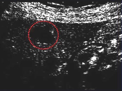 |
| CAD correctly classifies a well-differentiated HCC in an MFI image acquired using the Sonozoid contrast agent. All images courtesy of Junji Shiraishi, Ph.D. |
Microflow imaging is a five-step process that begins with normal B-mode ultrasound imaging to identify the location of the focal liver lesion, Shiraishi explained. The microbubble contrast agent is then injected for the harmonic imaging phase. The contrast agent is enhanced by use of a phase-inversion imaging method. Next, the burst scan clears the microbubbles within the scanning volume by use of high mechanical index scanning. In the last step, harmonic imaging with maximum-hold processing captures the replenishment of the microbubbles in the volume.
"Usually the [harmonic] image is obtained in less than 10 seconds because the patient needs a breath-hold," Shiraishi said.
All images were acquired on an Aplio XG ultrasound scanner (Toshiba) using AVI format, an acoustic frame rate of 15-30 frames per second, and a pixel size of 0.1-0.5 mm.
The CAD scheme processes the images in five major steps, beginning with extraction of the MFI data from the original cine clip. There are 25 extracted features in all, including morphological features, grayscale features, and temporal features such as time-intensity curve, derived from the MFI data, Shiraishi said.
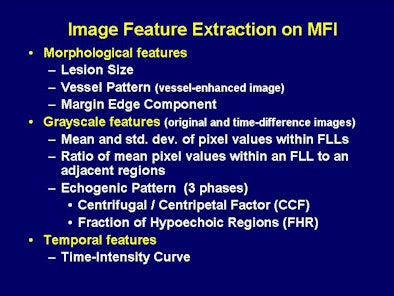 |
| Twenty-five image features are extracted from the MFI data for analysis by artificial neural networks (ANNs). |
The second step is segmentation of the organs including the kidney, vessels, and a region of the liver parenchyma. Then comes assessment of temporal, morphologic, and gray-level image features, followed by the application of six artificial neural networks (ANNs) based on the 25 image features.
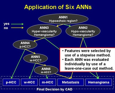 |
| Six independent ANNs analyzed the 25 microflow image features to distinguish HCC, metastases, and hemangioma. |
"Then we extracted the image features from the artificial neural networks to obtain estimation of likelihood measures for all five types of FLLs," including well-differentiated HCC (w-HCC), moderately differentiated HCC (m-HCC), and poorly differentiated HCC (p-HCC), as well as metastases and hemangiomas, he said.
A novel distinguishing feature in the grayscale analysis is known as centrifugal/centripetal factor (CCF).
"Centrifugal indicates echogenic enhancement from center to the periphery of the lesion," a pattern indicative of HCC, he said. "Centripetal indicates enhancement from the peripheral region to the center," suggestive of a hemangioma. The results of CCF analysis are used as a weighting factor by the ANNs.
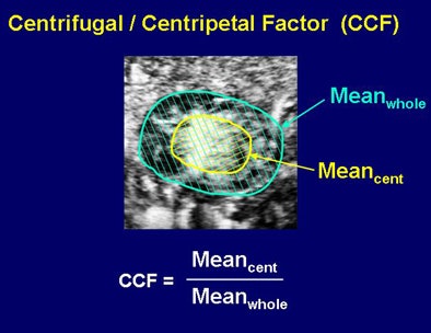 |
| Centrifugal/centripetal factor (CCF) analyzed whether contrast enhancement occurred from the periphery toward the center of the lesion (hemangioma) or from the center toward the periphery (HCC). |
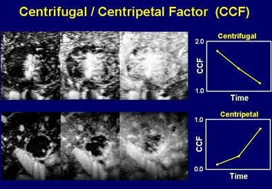 |
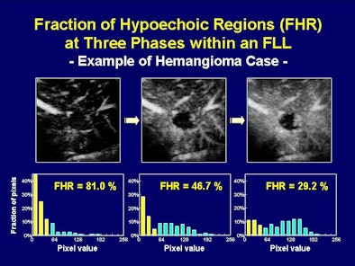
Another grayscale feature, fraction of hypoechoic regions (FHR) analysis, quantifies the percentage of hypoechoic regions in three phases, revealing distinct patterns for hemangiomas versus tumors and metastases.
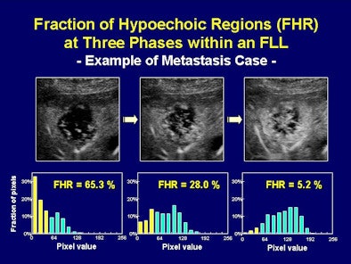 |
| The CAD algorithm analyzed the FHR at three phases within a focal liver lesion. Metastasis (above) and hemangioma (below) reveal different FHR profiles. |
 |
The retrospective study analyzed 146 focal liver lesions in 137 patients. There were a total of 76 histology-proven HCCs, including 24 well-differentiated HCCs, 36 moderately differentiated HCCs, 16 poorly differentiated HCCs, 40 metastatic lesions, and 30 hemangiomas. Hemangioma diagnoses were confirmed using contrast-enhanced CT or MRI.
Temporal features such as contrast accumulation times at early and delayed phases, flow rate, and estimated peak time were determined by the use of estimated time-intensity curves for FLL.
For example, based on research by Huang-Wei and colleagues (Investigative Radiology, March 2006, Vol. 41:3, pp. 363-368) the group used quantitative parametric analysis to differentiate focal nodular hyperplasia from other hypervascularized liver focal lesions.
The researchers applied six independent artificial neural networks to the 25 image features to classify five types of liver disease: metastases, hemangiomas, w-HCC, m-HCC, and p-HCC.
The classification accuracies for the 146 focal liver lesions were 67.5% for metastases, 80% for hemangiomas, 79.2% for well-differentiated HCCs, 75% for moderately differentiated HCCs, and 87.5% for poorly differentiated HCCs, Shiraishi said.
When applied to all three types of focal liver lesions (HCC, metastasis, and hemangioma), the classification accuracy for all HCCs was 94.7%. Conversely, the CAD scheme incorrectly classified 23 focal liver lesions (four HCCs, 13 metastases, and six hemangiomas) into the three broader types of liver disease. The overall classification accuracies for malignant (HCC and metastases) and benign (hemangioma) lesions were 97.4% and 80%, respectively, he noted.
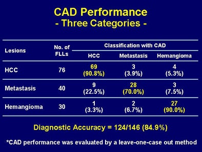 |
| CAD demonstrates high sensitivity for distinguishing three liver diseases (above); sensitivity varies when HCC lesions are classified as well-differentiated, moderately differentiated, or poorly differentiated. |
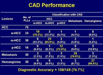 |
Compared to the group's previous study of 103 FLL using the SonoVue contrast agent, Sonozoid performed similarly, if slightly less effectively, in a database of 146 focal liver lesions. The CAD system's accuracy for distinguishing the three types of liver disease (HCC, metastasis, and hemangioma) was 88.3% for SonoVue versus 84.9% for Sonozoid. CAD accuracy for distinguishing five types of FLLs (well, moderately, and poorly distinguished HCC; metastasis; and hemangioma) was 75.7% for SonoVue and 74.7% for Sonozoid.
"Our CAD scheme for classifying FLLs has a potential to improve diagnostic accuracy in histological differential diagnosis of HCC and other liver diseases when CEUS images were obtained by use of MFI technique," Shiraishi concluded.
In response to a question from the audience, Shiraishi said that lesion size did not appear to affect the CAD system's classification accuracy in the study.
By Eric Barnes
AuntMinnie.com staff writer
September 29, 2008
Related Reading
Breast ultrasound CAD performance varies in ethnic population, September 5, 2008
CAD steals show from U.S. research database, August 12, 2008
Percutaneous radiofrequency ablation effective for small liver tumors, January 4, 2008
CEUS performs well with cystic pancreatic masses, November 23, 2007
Ablation for local HCC treatment on the rise, study finds, January 30, 2007
Copyright © 2008 AuntMinnie.com


