You know you're onto something when the big boys come calling. GE Healthcare has announced its acquisition of automated breast ultrasound (ABUS) developer U-Systems, a move that highlights the rapid evolution of ABUS from a niche technology into a promising adjunct to screening mammography.
U-Systems flagship product is somo.v, an ABUS system that automatically scans a woman's breast and displays images for review by a physician. ABUS proponents believe the technology is superior to freehand breast ultrasound, as it is less operator-dependent and is easier to perform from a workflow perspective.
GE plans to incorporate the ABUS technology into its ultrasound business, with distribution of somo.v handled by both the company's ultrasound and women's imaging units, according to Brian McEathron, general manager of general imaging ultrasound for GE.
"This will be part of the suite of breast care tools that GE offers, along with workflow products for breast care labs," McEathron told AuntMinnie.com. "This is another important piece of the puzzle to screen, diagnose, and treat women for breast cancer."
The company will retain both the somo.v and U-Systems brands, and it will maintain U-Systems facilities in California and Arizona. Financial terms of the deal were not disclosed.
1st to market
Somo.v in September 2012 became the first ABUS system to receive approval for use in a screening environment when the U.S. Food and Drug Administration (FDA) approved U-Systems' premarket approval (PMA) application. U-Systems already had 510(k) clearance for the product for diagnostic use, but the new approval expanded its application to the much broader screening market.
Technologies such as ABUS are of great interest to breast imaging specialists looking for tools to complement mammography in women with dense breast tissue, which confounds x-ray-based imaging techniques. Clinical studies that U-Systems submitted to the FDA in support of the PMA found that the use of somo.v along with mammography resulted in the detection of 30% more cancers than mammography alone.
 |
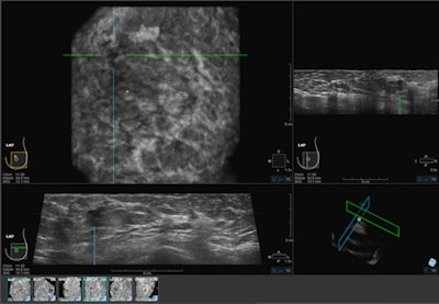
| ABUS image showing mass in upper-outer quadrant of the left breast. Image courtesy of U-Systems. |
Several studies on ABUS will be presented at the 2012 RSNA meeting, including one led by Dr. Rachel Brem of George Washington University Medical Center. Brem's study found that ABUS enabled detection of early-stage cancers in women with dense breasts, giving healthcare providers time to start early treatment. In all, 88% of cancers found by ABUS alone in a group of 15,000 women were grade 1 or 2.
Another study, to be presented by Maryellen Giger, PhD, of the University of Chicago, found that adding ABUS to mammography for women with dense breast tissue improved sensitivity by 23.3 percentage points, from 38.8% for mammography alone to 63.1% for mammography plus ABUS.
Business opportunity
All this growing clinical evidence has translated into a business opportunity for GE. Ultrasound long ago carved out a problem-solving role as an adjunct to mammography for diagnostic applications, but its use in screening was held back by the workflow challenges involved in performing freehand ultrasound on such a large number of patients.
Freehand ultrasound takes 45 minutes to perform, compared with only about 15 minutes for ABUS, according to McEathron. Also, freehand ultrasound must be performed by experienced personnel, while staff can be trained to position the ABUS system after just a two-day course.
ABUS images are still read by radiologists, but even that is easier than with freehand ultrasound. ABUS exams are typically presented as thick volumes six images at a time, and readers can drill into the studies for more information, McEathron said.
"You can use handheld ultrasound, but it's a long process, it takes a lot of physician time to perform, and it's not conducive to a screening environment," McEathron said. "With U-Systems, they have the workflow developed such that it's very efficient."
Rising awareness
Another factor leading to the acquisition has been increasing awareness of the importance of breast density in determining a woman's risk of developing breast cancer. A 2007 article in the New England Journal of Medicine found that women with dense tissue in more than 75% of the breast have a four to six times greater risk of cancer than women with the least dense tissue.
The situation has even become a political football, giving rise to a grassroots advocacy movement that seeks to require healthcare providers to notify women of their density status when they receive a mammogram. Starting in Connecticut and spreading to New York, Texas, Virginia, and California, the movement has been successful in getting density notification laws passed.
The movement hasn't gone unnoticed at GE, according to McEathron.
"I think the grassroots efforts in many states and advocacy groups trying to help women understand the issue are very important factors in the creation of this new clinical space, and having that advocacy start to develop is an important part of why we got into it," he said.
And with GE now backing ABUS, odds are that the technology will find its way into many more breast imaging clinics than if U-Systems had gone it alone.
"Eventually this will save a lot of lives collectively, between the awareness of what's going on and the fact that there is a tool available for women" with dense breast tissue, McEathron said.


