Thanks to advances in automation and improved imaging capabilities, B-mode ultrasound can be used to assess subclinical atherosclerotic disease and better identify those patients who would benefit from medical intervention prior to symptom onset, according to research published in Global Heart.
For the study, a multi-institutional, multinational research team examined two cohorts from India with automated B-mode ultrasound and compared the results with those of two cohorts from North America. Automated ultrasound of the carotid and iliofemoral arteries could feasibly provide rapid screening for subclinical atherosclerotic disease in a range of settings, the researchers found.
They also concluded that adding B-mode examination of the iliofemoral arterial beds to carotid ultrasound screening identifies additional subjects who would benefit from prophylactic medical intervention for atherosclerotic cardiovascular disease (ASCVD)-related events.
"Surely, such a simple approach merits adoption on a wide scale as a modern approach to a modern scourge of rapidly rising ASCVD-related events worldwide," wrote the authors, led by Ram Bedi, PhD, of the University of Washington's department of bioengineering, and senior author Dr. Jagat Narula, PhD, from Icahn School of Medicine.
Asymptomatic subjects
Doppler-based ultrasound systems are commonly used to assess carotid stenosis in symptomatic patients to identify those who would benefit from surgical intervention. The researchers believed that recent image quality improvements and advances in automation could enable B-mode systems to be similarly used for assessing subclinical atherosclerosis in the asymptomatic population to determine subjects who would benefit from prophylactic medical intervention (Global Heart, December 2014, Vol. 9:4, pp. 367-378).
To test their theory, the researchers used B-mode ultrasound to calculate the prevalence of atherosclerotic disease in 941 asymptomatic volunteers (mean age, 44.27 ± 13.76 years) from two underserved communities in India where ASCVD risk factor information was unknown. While one community was from an urban city (Jaipur), the other community -- from the semiurban town of Sirsa -- consisted of devout followers of a local spiritual leader, and the subjects had undergone aggressive lifestyle changes.
The results were compared with reference data gathered from two primary care clinics in North America: one was in Toronto, and the other was in Richmond, TX. The 481 subjects in this part of the study had a mean age of 59.68 ± 11.95 years; most were office workers in mid- to high-income brackets and few engaged in regular physical activity, according to the researchers.
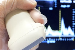
Automated bilateral B-mode ultrasound studies of the carotid and iliofemoral arteries were performed at two health camps in India, using a CardioHealth Station (Panasonic Healthcare) ultrasound system, by one of eight radiology residents. The residents did not have prior experience in vascular ultrasound but received two hours of training. Ultrasound exams were performed by trained vascular sonographers at the two North American clinics in the study.
While conventional 2D imaging was considered satisfactory for identifying focal lesions, 3D imaging data for the arterial segment of interest were acquired to automate the process of plaque identification and quantification. To present the clinical findings in an easy-to-understand manner, the researchers developed an index called the Fuster-Narula (FUN) score, which summarizes the intima-media volume for the scanned peripheral arteries.
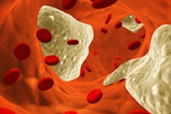
The researchers found that 224 (24%) of the participants from India had plaque in at least one of the four arterial sites. Furthermore, 107 (11%) had plaque only in the carotid arteries, 70 (7%) had plaque in both the carotid and iliofemoral arteries, and 47 (5%) had plaque only in the iliofemoral arteries. The presence of plaque was associated with older age and the male gender, but not with systolic blood pressure, the group noted.
Better treatment matching
In the North American clinics, 203 (42%) of the subjects had carotid plaque. Notably, 82% of these individuals would not have qualified for lipid-lowering therapy under the Adult Treatment Panel (ATP) III guidelines, and 33% would not have qualified for treatment under the recently published ATP IV Guidelines.
The ultrasound findings could also prevent overtreatment: 34 subjects from the North American clinics did not have plaque but qualified for treatment under the ATP III guidelines. In addition, 81 participants qualified for treatment under the ATP IV guidelines but did not have plaque.
"Our results showed that ultrasound examination, when compared with ATP III and ATP IV guidelines, allowed improved identification of individuals who could be targeted for prophylactic medical intervention," the authors wrote.
Although 2013 guidelines from the American College of Cardiology and the American Heart Association recommend against the use of carotid intima-media thickness testing, the panel that drafted the guidelines did not consider plaque findings, according to the researchers.
"Clearly, direct evidence of subclinical atherosclerotic plaque burden incorporated in a simplified and standardized FUN score index provides motivation for aggressive medical intervention," the authors wrote. "In developing countries such as India where a much younger population is afflicted with the devastating consequences of ASCVD-related events, an alternative strategy for identifying high-risk individuals may not be out of place."
"The lower incidence of plaque presence in a more-disciplined cohort reported here should inspire the promotion of cardiovascular health by lifestyle modification," the researchers also noted.


