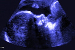
Female pelvic pain is often evaluated with MRI and CT, adding cost and -- in the case of CT -- radiation exposure for patients. Ultrasound is the better initial choice, according to an article published online Tuesday in the American Journal of Obstetrics & Gynecology.
Thanks to technological advances such as volume imaging, real-time transvaginal ultrasound (TVUS), and 3D Doppler techniques, ultrasound is ideally suited for evaluating the female pelvis and is an effective first-line imaging modality for most gynecologic patients, concluded a multi-institutional research team led by Dr. Beryl Benacerraf of Harvard Medical School.
"Consistent use of ultrasound imaging first in women with pelvic symptoms, especially with the adjunct of 3D and Doppler if necessary, would likely render further imaging unnecessary and at the same time be cost-effective and safer," the authors wrote in their Clinical Opinion article (Am J Obstet Gynecol, March 31, 2015).
Ultrasound First initiative
The American Institute of Ultrasound in Medicine's (AIUM) Ultrasound First initiative is particularly relevant for obstetric and gynecologic patients, "for whom a skillfully performed and well-interpreted ultrasound image usually obviates the need to proceed to additional more costly and complex cross-sectional imaging techniques," according to the authors.
"Yet still today, many women with pelvic pain, masses, or flank pain first undergo CT scans and those with Müllerian duct anomalies typically have MRIs," they wrote. "Not uncommonly, CT or MRI of the pelvis often yields indeterminate and confusing findings that then require clarification by ultrasound imaging."
3D/4D ultrasound is among the key technical advances that have sparked the modality's improved utility in gynecological imaging. While ultrasound used to require filling a woman's bladder and obtaining a series of 2D images one at a time, that's no longer the case.
"Today, 3D volume ultrasound imaging allows the automated acquisition of an entire volume that, in turn, can generate hundreds of images and be used to reconstruct any view in any orientation," the authors wrote. "Furthermore, 3D ultrasound imaging is less expensive and less time-consuming than MRI."
Reconstructed views of the pelvis -- such as the coronal view of the uterus -- have greatly improved the modality's ability to answer the vast majority of clinical questions in gynecology, according to the authors. For example, 3D volume sonography has been shown to be just as effective as MRI for demonstrating Müllerian duct anomalies.
3D ultrasound has also "emerged as the ideal imaging modality not only when examining patients with infertility but also for examining patients with pelvic pain associated with embedded intrauterine devices, fibroid tumors, adenomyosis, adnexal masses, torsion, [and] endometriosis," they wrote. "Ultrasound volume imaging makes it possible to localize fibroid tumors, polyps, and hydrosalpinges with high precision, as well as other uterine abnormalities."
As a result, the medical community must be educated to consider using 3D ultrasound for the initial assessment of specific gynecologic indications, such as evaluating the uterus for Müllerian abnormalities and localizing intrauterine devices or other intracavitary lesions, according to Benacerraf and colleagues.
"In this setting, it is likely that fewer women would require a costly workup that involves multiple advanced imaging studies if 3D ultrasound images were performed first," they wrote.
Real-time TVUS
The transvaginal transducer -- one of the most important innovations in pelvic imaging over the past few decades -- along with the real-time nature of ultrasound imaging enables close probing of pelvic organs to elicit patient symptoms and to correlate them with specific pelvic anatomic locations, according to the authors.
"The practitioner therefore can gain crucial information about the degree and area of pain and mobility of organs in the pelvis and correlate the ultrasound findings with the physical examination," they wrote. "The ability to examine and image the patient at the same time offers considerable and too often neglected value, which is unique to ultrasound imaging as a cross-sectional imaging technique."
For example, tenderness-guided ultrasound imaging is now the most effective way to detect implants of painful, deep-penetrating endometriosis throughout the pelvis, according to the group.
"Ultrasound imaging has proved to be accurate for the evaluation of deep infiltrating endometriosis and for patients with pain because of extensive pelvic adhesions," they wrote. "Not only can we identify abnormalities on the images, but also simultaneous gentle pushing can show whether organs slide past each other, thus providing crucial information about the origin of a mass (adnexal mass versus broad ligament fibroid tumor) and the connections and adhesions between the organs."
Real-time ultrasound also facilitates sonohysterography, in which a small catheter is used to inject saline solution into the uterine cavity. Clinicians can then evaluate the endometrium for polyps, submucosal fibroid tumors, synechiae, and uterine shape, according to Benacerraf and colleagues.
"Adjunctive to sonohysterography, the installation of microbubbles is useful for the determination of tubal patency and is critical to those patients with contrast allergies," they wrote.
3D Doppler for blood flow
Ultrasound combines morphologic and vascular imaging, an advantage for characterizing pelvic masses. Without requiring the injection of contrast, Doppler yields valuable information about the location and degree of blood flow in and around pelvic lesions, according to the authors.
"Not only is the characteristic grayscale image of pelvic abnormalities key in making a diagnosis, but also 3D Doppler ultrasound imaging can evaluate the mapping and density of blood flow and even provide a quantitative measure of the amount of blood flow in a lesion," they wrote.
Color Doppler mapping also provides important information for evaluating adnexal masses and differentiating an endometrioma from an ovarian tumor or an ovarian fibroma.
"For example, the unique Doppler pattern of a hemorrhagic corpus luteum permits this definitive diagnosis as a cause of acute pelvic pain," they wrote.
An education gap?
The authors noted that it would be best if all practitioners could be comfortable with high-resolution 3D ultrasound imaging, tenderness-guided TVUS, and pelvic Doppler imaging.
"It is unfortunate that ultrasound users have such a wide range of experience, such that not everyone uses the modality to its full potential," they wrote. "Inexperience should not justify ordering an MRI or CT scan."
Although ultrasound technology has advanced very quickly, many practitioners still provide only basic 2D ultrasound imaging. This dynamic highlights the need for education and dissemination of information on the newer techniques that ultrasound offers, according to the authors.
"In this era of cost concerns, it is very important to recognize that ultrasound technology now offers multiple applications such as 3D volume imaging (similar to CT and MRI), real-time evaluation of pelvic organs along [with] the physical examination, and Doppler blood flow mapping (without contrast)," they wrote. "Collectively, these applications make ultrasound imaging a unique imaging modality that ideally is suited to evaluate the female pelvis."


