Contrast-enhanced ultrasound (CEUS) is highly effective for characterizing tumors thought to be hepatocellular carcinoma (HCC) but indeterminate on MRI, according to a presentation at the 2016 Advances in Contrast Ultrasound - Bubble Conference earlier this month in Chicago.
Sharing preliminary findings from an ongoing study, senior author Dr. Stephanie Wilson of the University of Calgary reported on her institution's experience using CEUS to resolve more than 50 cases so far with suspicion for HCC and indeterminate findings on MRI. Wilson said she hopes that such studies will facilitate the integration of CEUS into existing imaging guidelines for liver imaging.
"I hope that after every place on the guidelines that currently says, '[after an] indeterminate MR, do a CT scan' or 'do biopsy,' it will say instead, 'do CEUS,' " she said.
MRI as a mainstay
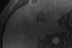
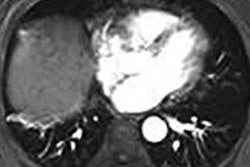
MRI remains the mainstay of imaging for hepatocellular carcinoma in 2016, both in North America and in other parts of the world, Wilson said. However, MRI can also produce an indeterminate result due, for example, to the absence of the required contrast-enhancement features for an HCC diagnosis: arterial phase hyperenhancement (APHE) and slow contrast washout.
An indeterminate result may also occur due to the lack of baseline observations on T1-, T2-, and diffusion-weighted imaging (DWI), Wilson said. The American Association for the Study of Liver Diseases (AASLD) recommended in 2011 that indeterminate results on MRI be considered for biopsy.
CEUS can help, however. The University of Calgary research team, which included first author and medical student Jingui Hui, PhD, performed a prospective study, recruiting all patients who were considered to be at risk for HCC and had temporally related CEUS and MRI exams. In a six-month period from July 2015 to January 2016, 102 patients were enrolled in the study, which is ongoing. Data analysis is still in progress for the initial six-month period, but Wilson shared preliminary results in her talk in Chicago.
Of the 102 patients, 52 (51%) had an MRI report that suggested an indeterminate result. This included 49 patients with an abnormal parenchymal observation; 35 of these had a total of 41 mass-like observations, including 26 mass-like observations that showed arterial phase contrast hyperenhancement but not portal venous-phase washout on MRI.
In addition, 14 patients with an abnormal parenchymal observation had arterial phase hyperenhancement only, without a baseline T1, T2, or DWI abnormality and without contrast washout; these observations were suspected to be arterial portal shunts due to their appearance, Wilson said. The remaining three patients with indeterminate results on MRI had an indeterminate thrombus in the portal vein.
Resolution with CEUS
Of the 26 patients with MR findings of a mass-like area with arterial phase hyperenhancement only, 12 had a nodule on the baseline ultrasound scan that was not seen on MRI. Eleven of the 12 had classic HCC features on CEUS: arterial phase hyperenhancement and weak late contrast washout, she said. The other patient had classic nonhepatocellular carcinoma features on CEUS: arterial phase hyperenhancement and rapid contrast washout.
Five cases are biopsy-proven for HCC, including four of HCC and the one case of nonhepatocellular carcinoma, which was confirmed as cholangiocellular carcinoma (CCC).
"All of these [12 cases] have been treated as cancer," she said.
CEUS can also be an excellent method for resolving cases of suspected arterial portal shunts, according to Wilson.
Among the 14 cases with suspected arterial portal shunts, 13 had completely negative findings on CEUS, with no baseline nodule, hyperenhancement, or washout.
"So this would support that these are correctly identified as arterial portal shunts," she said.
However, CEUS did show a nodule in one case that had classic enhancement features for HCC, Wilson said.
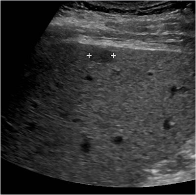 Above, small superficial hypoechoic mass (marked by calipers) in the cirrhotic liver of a 64-year-old man. Below, at 17 seconds after contrast injection, the mass is hypervascular relative to the remainder of the liver and appears brighter on this image. All images courtesy of Dr. Stephanie Wilson.
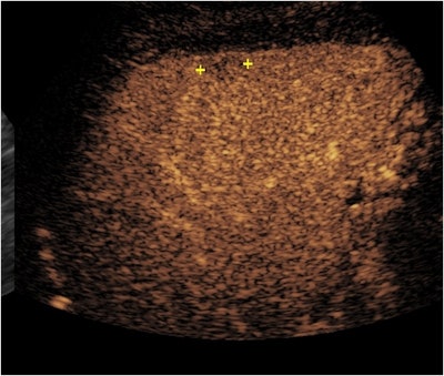 Above, at one minute and 36 seconds after contrast injection, the mass (marked by calipers) shows very faint reduction in enhancement and washout. Below, at four minutes and 35 seconds after contrast injection, the mass is now showing more marked washout. The arterial phase hyperenhancement and slow washout shown in this CEUS study are classic features of HCC.
CEUS can also differentiate between bland and tumor thrombus. This is very important, as the presence of tumor thrombus significantly changes cancer staging, treatment options, and prognosis, she said. Of the three patients in the study with an indeterminate thrombus in the portal vein, CEUS found that one patient had two bland thrombi and two patients each had a tumor thrombus.
"[CEUS] has been shown to be the most sensitive modality for detecting neovascularity within a tumor thrombus," she said.
Adding CEUS to guidelines
Wilson also hopes that the imminent release of new AASLD guidelines will incorporate CEUS, which had been removed in 2011. In other positive developments, the initial publication in June of the American College of Radiology's Liver Imaging Reporting and Data System (LI-RADS) for CEUS now accompanies the already existing LI-RADS publication for CT and MRI, she said.
"This is a huge advantage for CEUS and will pave the way for CEUS advancement in the U.S. and elsewhere," she said.


