
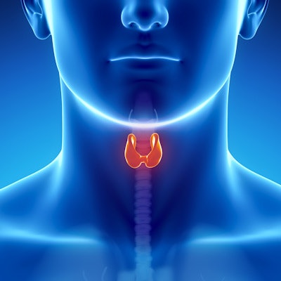
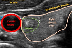
Although it doesn't perform as well as a sonography expert, ultrasound computer-aided diagnosis (CADx) software can still be useful for stratifying thyroid nodules by their risk of malignancy, according to research published online April 11 in the American Journal of Roentgenology.
A team of researchers led by Dr. Jordi Reverter, PhD, of the Autonomous University of Barcelona in Badalona, Spain, found that a commercially available ultrasound CADx software application produced similar sensitivity and negative predictive value to an experienced endocrinologist in analysis of 300 thyroid nodules. It did, however, have lower specificity and positive predictive value.
"Given the high sensitivity and [negative predictive value] that we reported (at least for preliminary screening procedures), such systems could conceivably allow image classification of thyroid nodules as benign or malignant to be performed by a nonexpert operator such as a technician, although the final risk classification should be made by an experienced professional," the authors wrote.
Seeking to assess the performance of the AmCAD-UT software (AmCad BioMed) as a diagnostic aid, the researchers retrospectively gathered ultrasound images of 300 thyroid nodules in patients from the endocrinology service of their large public teaching hospital. Of the 300 nodules, 135 were malignant. The mean maximum nodule diameter was 3.2 ± 1.0 cm for malignant nodules and 2.8 ± 0.4 cm for benign nodules.
A senior staff endocrinologist with 10 years of experience in diagnostic and therapeutic thyroid ultrasound procedures reviewed all of the images and assigned each a suspicion of malignancy score based on the American Thyroid Association (ATA) system. The CADx software was then applied to categorize the images based on the ATA system, the European Thyroid Imaging Reporting and Data System (EU-TIRADS), and the system jointly proposed by the American Association of Clinical Endocrinologists (AACE), the American College of Endocrinology (ACE), and the Associazione Medici Endocrinologi (AME). The researchers then compared the CADx software's results with the endocrinologist's ratings.
| Performance for classifying risk of thyroid nodules | ||||
| CADx software based on AACE/ACE/AME classification system | CADx software based on EU-TIRADS classification system | CADx software based on ATA classification system | Clinical expert using ATA classification system | |
| Sensitivity | 81.5% | 85.2% | 87% | 87% |
| Specificity | 53.2% | 50.2% | 68.8% | 91.2% |
| Positive predictive value | 51.8% | 50.1% | 64.5% | 90.5% |
| Negative predictive value | 80.8% | 82.6% | 86.3% | 90.9% |
| Area under the curve | 0.70 | 0.71 | 0.72 | 0.88 |
The CADx software performed best when applying the ATA reporting system, yielding statistically similar sensitivity and negative predictive value to the clinical expert. However, its specificity and positive predictive value were significantly lower (p < 0.01).
"The fact that an expert would achieve superior results is unsurprising, given that the CADx system learns from the accumulated expertise of imaging experts, which is stored in its database," the authors wrote.
The researchers concluded, however, that the software was reliable and easy to use, making it useful in clinical practice.






![Examples of ultrasound findings and techniques. (A) Images in a 39-year-old male patient with a mass in the left thigh. The mass is heterogeneous on the B-mode US image (compared with the patient in D) and showed increased microvascularity (superb microvascular imaging [SMI]) and shear-wave elastography (SWE) values. Undifferentiated pleomorphic sarcoma was diagnosed at biopsy (with pleomorphic rhabdomyosarcoma in surgical specimen). (B) Images in an 18-year-old male patient with a mass in the left leg. The mass is hypoechoic on the B-mode image, with no other findings suggestive of malignancy. The lesion is in contact with the cortex of the tibia, which is slightly irregular. CT revealed a doubtful anteromedial tibial erosion. The microvascular study demonstrated high vascularization, suggestive of malignancy. Periosteal Ewing sarcoma was diagnosed with both histologic and immunohistochemical confirmation. (C) Images in a 69-year-old female patient with a lump growing on the outside of the left leg. Multiple SWE examinations were performed (please note the high values obtained in the measurements, whereas the color map highlights the stiffness relative to adjacent tissues). SMI showed areas of increased vascularization to target for sampling. Undifferentiated spindle cell sarcoma was diagnosed at biopsy, with residual leiomyosarcoma in the surgical specimen after neoadjuvant therapy. (D) Images in a 56-year-old female patient with a mass in the right thigh. The mass is heterogeneous at both B-mode ultrasound (similar to patient A) and MRI (coronal T2-weighted spectral attenuated inversion recovery [SPAIR]; T1-weighted pre-contrast and postcontrast imaging), which even shows uptake after the administration of paramagnetic contrast material, which is traditionally suggestive of malignancy. Low values at SMI and elastography are suggestive of benignity. Spindle cell lipoma was diagnosed at biopsy, with atypical spindle cell lipomatous tumor in the surgical specimen.](https://img.auntminnie.com/mindful/smg/workspaces/default/uploads/2026/08/images-radiol250278fig2.APCFLSvX6p.jpg?auto=format%2Ccompress&dpr=2&fit=crop&h=167&q=70&w=250)
