Point-of-care ultrasound (POCUS) has unique strengths that make it promising for evaluating broken bones in children. But could POCUS really replace x-rays for fractures? The answer may depend on training, according to the findings of an October 21 study in Ultrasound in Medicine and Biology.
Researchers from Italy and the U.S. compared the performance of emergency physicians using POCUS to diagnose pediatric fractures to that of radiologists identifying fractures on radiographs. While the overall performance of POCUS was subpar, some highly experienced operators achieved results comparable to radiologists.
"Providers with greater skill performed significantly better than those with a standard skill level, suggesting that more training and experience is required for pediatric fracture detection," wrote the authors, led by Dr. Constantino Caroselli, a geriatric emergency physician from Ancona, Italy.
The pilot study included 547 children under the age of 18 with suspected limb, hand/feet, or thorax fractures. The children visited one of five medical centers in Italy, where they underwent imaging with both POCUS and radiographs.
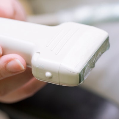
One trained emergency physician at each institution performed the point-of-care scans with a high-frequency linear transducer in both the longitudinal and transverse orientations. Meanwhile, trained radiologists read the patients' radiographs, which were taken with portable or fixed equipment.
| Performance of emergency physicians using POCUS to diagnose fracture in children | |||
| All physicians | Standard-skill physicians | Highly skilled physicians | |
| Sensitivity | 74.5% | 71.5% | 91.7% |
| Specificity | 83.6% | 82.9% | 88.9% |
| Agreement with x-ray | 0.58 | 0.54 | 0.81 |
The x-ray results, which served as the reference standard, identified fractures in 43% of the participants. The overall performance of all five emergency physicians netted a sensitivity of 74.5% and 83.6%, and the agreement coefficient was 0.58, demonstrating only moderate alignment between POCUS and x-ray findings.
However, the results varied greatly among the five emergency physicians. Physicians with a high level of POCUS experience, defined as at least 18 months of training and 200 supervised exams, had excellent agreement with x-rays. In fact, the best-performing provider achieved a 100% sensitivity and specificity.
The study reinforces that the performance of POCUS is highly dependent on the skillset of the operator. It also reflects issues around POCUS training, licensing, and credentialing -- topics increasingly talked about in the radiology and ultrasound communities.
If medical institutions can achieve consistently excellent results with POCUS, it could be a boon to emergency departments for pediatric fractures, the authors noted. POCUS does not use ionizing radiation, can detect soft-tissue injury, and is associated with less pain. It would also eliminate the time delay between when a patient is examined and when they get their results.
"A POCUS examination of bones is brief, painless, and relatively simple to perform -- all ideal qualities for the rapid pace required in emergency departments," the authors wrote.
While the benefits of POCUS are clear, the authors said more studies are needed to address some of the larger training and privileging questions.
"Further study determining the exact learning curves among novices to advanced POCUS users would help determine an appropriate training cut-off for pediatric fracture evaluation with ultrasound," they concluded.