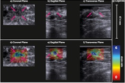
Microflow imaging can differentiate between benign and malignant breast lesions, suggest findings published October 28 in Clinical Radiology.
Researchers led by Runlan Luo, MD, from the Chinese PLA General Hospital in Beijing found that this method along with high-definition microflow imaging outperforms color Doppler flow imaging in diagnosing breast lesions.
“Microflow imaging and high-definition microflow imaging can detect more blood flow in breast lesions than color Doppler flow imaging…,” Luo and co-authors wrote. “High-definition microflow imaging could reduce the unnecessary biopsy of ultrasound BI-RADS 4A lesions without missed malignancy.”
Microflow imaging is a newly released Doppler ultrasonic imaging technology, in which intelligent filtering software can distinguish fine tissue movement from slow-flowing microvessels. The researchers highlighted that it could detect microvessels with an inner diameter of 0.1 mm without being angle-dependent and display microvascular patterns.
High-definition microflow imaging meanwhile has been touted as an advanced version of the original method. The researchers wrote that this could further suppress tissue motion artifacts and improve the display rate of microvessels.
Previous research suggests that microflow imaging has a higher sensitivity in detecting vessels compared with color Doppler flow imaging in hepatic tumors.
Luo and colleagues sought to test both versions of microflow imaging, comparing their respective performances with that of color Doppler flow imaging in breast lesions.
The researchers included data from 133 women with 138 breast lesions, of which 80 were benign and 58 were malignant. They evaluated penetrating vessels and blood flow signals that were graded into four types: grade 0 (no obvious blood flow signal), 1 (one or two small blood vessels with a diameter of less than 1 mm), 2 (three or four small blood vessels), and 3 (more than four blood vessels or a network of blood vessels).
The team found that high-definition microflow imaging detected more blood flow of grade 3 and outperformed color Doppler flow imaging.
| Comparison between flow imaging methods for detecting blood flow in breast lesions | |||
|---|---|---|---|
| Blood flow (Adler’s grade) | Color Doppler flow imaging | Microflow imaging | High-definition microflow imaging |
| Grade 0 | 29.7% | 9.4% | 6.5% |
| Grade 1 | 25.4% | 10.9% | 8% |
| Grade 2 | 23.9% | 15.9% | 12.3% |
| Grade 3 | 21% | 63.8% | 73.2% |
| *All data achieved statistical significance. | |||
The team also found that high-definition microflow imaging detected more penetrating vessels (31.9%) than the color Doppler method (17.4%, p = 0.018). However, the group also reported no significant difference between the two microflow imaging methods in detecting penetrating vessels.
The researchers also found that rich blood flow signals, penetrating vessels, and root hair-like and crab claw-like patterns were more likely in malignant breast lesions. Meanwhile, few blood flow signals and tree-like patterning were mostly in benign lesions, they reported.
Finally, the team found that microflow imaging could reduce unnecessary biopsy of 52 ultrasound BI-RADS 4A lesions with only two malignancies missed. Also, high-definition microflow imaging could downgrade 56 ultrasound BI-RADS 4A lesions with no malignancies missed.
The study authors suggested that microflow imaging, especially the high-definition version, can help detect breast disease earlier. They called for further in-depth studies to study the correlations between microflow imaging and microvessel density.
The study can be found in its entirety here.


