A medical team at the University of California (UC) Davis Health recently performed what it’s calling the world’s first endoscopic, ultrasound-guided core biopsy of a pancreatic tumor with a drilling method.
The team, led by Antonio Mendoza-Ladd, MD, said that this method successfully collected a larger-than-normal core of tumor tissue in an efficient manner. It highlighted that this can make way for better diagnostic workup.
“It is done in an easier-to-control way. We only need to do one puncture,” Mendoza-Ladd told AuntMinnie.com. “The cores we obtain with this device are better than regular needles.”
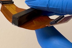
The medical team performed the procedure by using an instrument called EndoDrill GI (BiBB Instruments) to biopsy a gastrointestinal stromal tumor and two pancreatic tumors. The team highlighted that it could better access deep tissues in the upper gastrointestinal tract.
The technology uses electric high-speed drilling to take core tissue samples. An endoscopist operates a flexible rotating cylinder to remove fine samples while maintaining tissue architecture. The technology also enables sampling for deep-lying tumors. The rotating needle also removes fragments.
“It’s like a drill that you use at home for work you need to do around the house,” Mendoza-Ladd said, talking about the motion of the drill.
He added that this method avoids the need for a stabbing motion to be used to create the initial puncture in conventional biopsies.
Mendoza-Ladd said that the patients who underwent the procedure have not experienced post-procedure complications. He also highlighted that the samples from the procedure make way for more accurate diagnosis and ergo, better treatment strategies for gastrointestinal cancers.
“The hope is that we will reduce the number of punctures that we need to do,” Mendoza-Ladd told AuntMinnie.com. “We will have a single pass that will give us enough tissue we can run on special analysis.”


