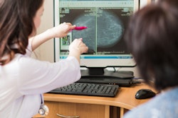
Combining radiomics and artificial neural network-based features improves ultrasound-based categorization of breast tumors compared with segmentation-based approaches, researchers have reported.
The findings could translate to more reliable ultrasound results by mitigating variations in operator skill, wrote a team led by doctoral student Zuzanna Anna Magnuska of RWTH Aachen University in Germany. The results were published September 10 in Radiology.
"Ultrasound is clinically established for breast imaging, but its diagnostic performance depends on operator experience," the group noted. "Computer-assisted (real-time) image analysis may help in overcoming this limitation."
Radiomics converts imaging data into quantifiable features and shows promise for facilitating ultrasound image-based breast cancer diagnosis, the authors explained. But using radiomics alone may not be the most effective way to categorize breast lesions with the modality, they wrote, noting that data on the efficacy of "direct mixing of various types of image-based features for breast tumor categorization on ultrasound images has not yet been reported."
To address the knowledge gap, Magnuska and colleagues explored whether combining classic and deep learning-based features would improve the sensitivity, specificity, and area under the receiver operating characteristic curve (AUC) of ultrasound-based radiomics for the categorization of benign and malignant breast tumors. They did so via a study that included data from 1,619 B-mode ultrasound images of breast tumors and used a tumor categorization model that combined radiomics and artificial neural network (also known as autoencoder) features (nnU-Net).
The study found the following:
- The model achieved an AUC of 0.9, sensitivity of 81%, and specificity of 87%.
- The study did not show any statistical difference between the model and human readers in breast tumor classification (AUC, 0.9 and 0.83, p = 0.55) or between the model and histopathologically or follow-up-confirmed diagnosis (AUC = 0.9 compared with 1; p = 0.1).
- The use of nnU-Net reduced the false-negative rate from 15% to 1% compared to a previous study conducted by the team.
"This study is, to the best of our knowledge, the first to use nnU-Net for breast lesion localization on B-mode ultrasound images," the authors wrote. "We showed that our lesion localization is reproducible [and] independent of added noise (p < 0.01 [and] p < 0.001)."
The research adds to existing literature regarding AI and radiomics for ultrasound, wrote Manisha Bahl, MD, of Massachusetts General Hospital in Boston, in an accompanying editorial.
"[The study] provides insights into how to build powerful segmentation and classification models," she noted.
The complete study can be found here.


![Delineation workflow: The various steps of the contouring process: (a) the nipple is manually annotated (b) a reference point is placed 30 mm behind the nipple (c) 10 random theta/displacement values are selected to obtain the central point of each ROI (d) Regions of interest are placed on the image. (e) The resulting 10 ROIs obtained for a representative subject (viewing window [0 10000]). Images are published under a Creative Commons license (CC BY-NC-ND 4.0).](https://img.auntminnie.com/files/base/smg/all/image/2025/02/1_s2.0_S1120179725000523_gr1_lrg.67bcbbc3ac5a9.png?auto=format%2Ccompress&fit=crop&h=167&q=70&w=250)