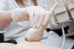
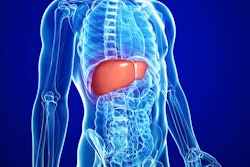
An ultrasound measurement technique could detect and stage acute cellular rejection in liver transplant patients, according to research published October 16 in Ultrasound in Medicine & Biology.
A group led by Luiz Vasconcelos, PhD, from the Mayo Clinic in Rochester, MN, found that the Attenuation Measuring Ultrasound Shear wave Elastography (AMUSE) technique has utility in this area, showing “excellent” agreement with biopsy-based diagnosis.
“These findings support the use of AMUSE potential for detection and staging of liver acute cellular rejection,” the team wrote.
Acute cellular rejection is an early post-liver-transplant complication that occurs in between 20% and 40% of patients. This is treated by corticosteroids. Another early complication in these patients is ischemia-reperfusion injury (IRI), which has similar laboratory results but is usually resolved without the use of therapy.
To distinguish between the two complications, liver biopsy is needed, which could cause further complications.
The AMUSE technique assesses shear wave velocity and attenuation. Previous studies suggest that AMUSE has the potential for noninvasive tissue characterization. Vasconcelos and colleagues previously demonstrated that AMUSE could distinguish between transplanted livers with and without acute cellular rejection.
For their current study, the authors studied AMUSE’s potential as a noninvasive alternative to differentiate acute cellular rejection from IRI. From there, they compared the results to that of the gold standard diagnosis of liver biopsy. In patients with cellular rejection, the team analyzed AMUSE’s ability to track response to corticosteroid therapy.
The investigation included data from 58 transplanted livers. Of the total patients, 13 underwent longitudinal tracking from cellular rejection diagnosis on Day 7 to therapy initiation and repeat biopsy on Day 14.
AMUSE measurements at 100 Hz, 200 Hz, and 300 Hz demonstrated statistical significance (p < 0.001 for all) for acute cellular rejection presence.
The team highlighted that the 200 Hz measurement showed the highest Spearman correlation coefficients for shear wave velocity and attenuation (0.68 and -0.83, respectively). Also, high shear wave velocity (> 2.2 m/s) and low attenuation (< 130 Np/m) at 200 Hz correlated with cellular rejection diagnostic, while low velocity and high attenuation indicated no cellular rejection.
Why 200 Hz was the magic number for AMUSE was not clear, they wrote.
“The evaluation of how different frequencies can interact with injured liver tissue is beyond the scope for this study,” the group noted. “Some possible explanations could be that the energy is highest at this frequency. Additionally, attenuation effects increase with frequency, so measurements at 300 Hz might be confounded by lower energy and therefore higher variability.”
Vasconcelos and colleagues also combined shear wave velocity and attenuation into a single biomarker, which resulted in an F1 score (which measures predictive performance on a 0-1 scale) of 0.97. This combination further improved patient differentiation, they wrote.
The authors also used a support vector machine (SVM) learning algorithm to evaluate AMUSE’s ability to stage acute cellular rejection. They found that AMUSE achieved an F1 score of 0.95 and an area under the receiver operating curve (AUROC) value of 0.99 for staging.
“When evaluating the presence of acute cellular rejection, the SVM reached 0.99 F1 score, with 1.00 sensitivity/recall,” they reported.
Vasconcelos and colleagues highlighted that with these results in mind, the AMUSE method could allow physicians to track changes in liver viscoelasticity more frequently as well as the response to treatment without biopsy.
The full results can be found here.


