An imaging AI tool developed by researchers in China appears promising for assisting novice sonologists with musculoskeletal (MSK) ultrasound exams, according to a study published June 9 in Radiology.
Led by Xinyi Tang, MD, from Sichuan University and colleagues, the research could lead to wider adoption of ultrasound for examining bones, muscles, and vascular structures -- anatomic structures typically evaluated using CT or MRI.
"The wrist–hand region presents unique challenges," Tang and colleagues noted. "The steep learning curve and the lack of standardized quality control mechanisms make it difficult for beginners and even experienced sonologists in resource-limited settings to ensure consistent high-quality [ultrasound] examinations."
To that end, the researchers constructed their deep learning model from 66,743 wrist–hand ultrasound scans (obtained from 430 healthy volunteers at 20 hospitals), among which key anatomic structures of approximately 40% of the standardized plane images were annotated.
The software was designed to perform two key tasks: standard plane recognition and automatic key anatomy segmentation. First, researchers highlighted the best-performing architectures for each: High-Resolution Network (HRNet) for anatomic identification accuracy, and ResNet-50 for standard plane recognition.
Next, the clinical evaluation phase involved 36 healthy volunteers who were randomly assigned to undergo AI-assisted or conventional scanning by three junior-level and three senior-level novice MSK sonologists.
According to the results, the tool achieved an F1 score of 96.2% in identifying 15 standard planes.
Researchers noted that AI assistance had no evidence of an effect on the overall time required to complete the acquisition of the 15 standard planes with respect to the conventional method, with a median of 305.5 seconds for the AI-assisted group and 304 seconds for the conventional group (p = 0.58).
However, for novice MSK sonologists, AI assistance offered benefits in reducing scan time (AI-assisted group, 288 seconds versus manual group, 356 seconds), according to the team.
Notably, the proportion of high-quality (grade A, exemplary) images increased from 80.7% (218 of 270) to 93.3% (252 of 270), representing a 1.2-fold increase, whereas the proportion of low-quality (grade C, unacceptable) images decreased from 10.7% (29 of 270) to 1.9% (five of 270), representing a 5.8-fold reduction in the acquisition of nonstandard images (adjusted p < .05 for both), Tang and colleagues found. They also observed the improvements in both senior-level novice and junior-level novice MSK sonologists.
With assistance from the AI system, the standardized plane acquisition rate for novice MSK sonologists reached 98.1%, a 9.9% improvement over conventional scanning.
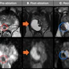
 AI-acquired standardized planes. Example ultrasound images acquired in clinical settings with the on-cart, AI-based software, illustrating automated acquisition and anatomic annotation of three standard planes: (A) S5 dorsal longitudinal plane of the wrist joint, (B) S11 transverse section of the proximal carpal tunnel, and (C) S14 transverse section of the second and third flexor tendons. A1 = trochlea A1, Cap = capitate, C-M.JS = recess of the capitate-metacarpal joint, D&Ss = flexor digitorum profundus and superficialis tendons, FCR = flexor carpi radialis tendon, FD = flexor digitorum tendon, FPL = flexor pollicis longus tendon, L-C.JS = recess of the lunate-capitate joint, Lun = lunate, MN = median nerve, Pis = pisiform, Rad = radius, R-C.JS = recess of the radiocarpal joint, Sca = scaphoid, UA = ulnar artery, UN = ulnar nerve, 3rd Met = third metacarpal.RSNA
The findings underscore the potential of the AI tool to standardize MSK ultrasound examinations and improve workflow efficiency, Tang and colleagues noted. Future work will expand to multiplatform, multipathology datasets, and large-scale trials to further evaluate the robustness and clinical value of the proposed AI model, they added.
In an accompanying editorial, Hospital of the University of Pennsylvania Chief of Musculoskeletal Imaging Levon Nazarian, MD, said hands-on ultrasound training in most radiology residencies has become less robust in recent years.
"The need for education is even more acute as MSK US grows outside the field of radiology," Nazarian wrote. If properly realized, the application of AI to MSK ultrasound can have far-reaching effects."
Increasing patient throughput by making MSK ultrasound more efficient could improve the economic bottom line and encourage radiology departments to support an MSK ultrasound service, Nazarian noted.
Access the full study here.