Dear AuntMinnie Member,
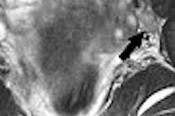
While the number of cervical cancer cases has declined steadily over the past 10 years, the disease is still far more prevalent that it ought to be. When cervical disease does develop, medical imaging can play an important role in managing patients, according to an article by staff writer Shalmali Pal that we're featuring in our Women's Imaging Digital Community this week.
The story describes how a group from Canada used dynamic contrast-enhanced MRI to detect changes in tumor vasculature that could provide a method of staging cancer. While the results were mixed, the researchers did find some parameters of contrast enhancement that could be valuable in tracking tumor behavior.
The article also profiles the work of a U.S. group that provided a new analysis of a study that compared CT and MRI in the pretreatment evaluation of cervical cancer, as well as research by a group from Italy on controlling the spread of cervical cancer to the lymph nodes. Get the entire story by clicking here.
In other women's imaging news, U.S. researchers look into why some patients have to undergo a second uterine artery embolization, in a story available by clicking here, while another group proposes a method for helping women avoid fibroids in the first place, available here.
Get these stories and more in our Women's Imaging Digital Community, at women.auntminnie.com.


