An MRI-based fusion radiomics model may offer non-invasive biomarkers and improve breast cancer management, according to research published June 1 in Magnetic Resonance Imaging.
A team led by Yuntai Cao, MD, from the Affiliated Hospital of Qinghai University in Xining, China found that its intratumoral-peritumoral radiomics fusion model, which used various MRI methods, achieved high diagnostic accuracy for assessing HER2-positive or negative breast cancers.
“The model not only provides reliable imaging biomarkers… but, more importantly, its predictive results can directly guide the selection of clinical treatment strategies, thereby advancing breast cancer diagnosis and treatment towards precision and individualized approaches,” the Cao team wrote.
Determining HER2 status is important for guiding proper treatment for breast cancer patients. Current clinical methods for assessing HER2 status are invasive, may have delayed results, and have limited sampling scopes.
Cao and colleagues explored how multiparametric MRI methods could help predict HER2 status in breast cancer. To do this, they integrated intratumoral and peritumoral radiomics features to establish a multiparametric MRI intratumoral and peritumoral radiomics model. This included using the following MRI techniques for the model: dynamic contrast-enhanced MRI (DCE-MRI), diffusion-weighted imaging (DWI), and T2-weighted fat-suppressed imaging (T2WI).
The team used a retrospective dataset of 266 women in its training and validation sets at two centers. Center one included 199 women who were divided into a training set (n = 140) and validation set (n = 59). Center two meanwhile included 67 women in an external test set.
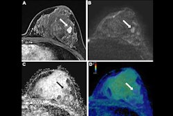
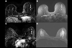
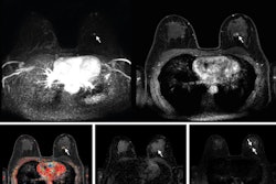
 The results depict tumor segmentation. an example of HER2 positive breast cancer is shown: (A), (B), and (C) represent the segmentation results of the entire tumor region on the T2WI, DCE-MRI, and DWI sequences, respectively; (D), (E), And (F) show the segmentation results of the intratumoral region combined with 3 mm peritumoral area on the T2WI, DCE-MRI, and DWI sequences, respectively.Available for reuse under Creative Commons license (Attribution 4.0 International CC BY 4.0 Deed)
The researchers used 3D Slicer to manually segment tumor boundaries on MRI exams to define intratumoral volumes of interest, which were expanded by 3 mm to generate peritumoral regions (VOI_Peri3mm). Finally, they extracted radiomics features to train eight random forest models.
Of the eight models, the multiparametric MRI intratumoral and peritumoral radiomics fusion model incorporating all three MRI techniques achieved the best HER2 prediction marks. It significantly outperformed single-parameter or single-region models.
Comparison between single-parameter, fusion MRI-based radiomics models | ||
Data set | Single-parameter model | Fusion model |
Training set | 0.739 | 0.822 |
Internal validation set | 0.754 | 0.823 |
External test set | 0.7 | 0.813 |
| *All data achieved statistical significance. | ||
The results point toward the model serving as a reliable imaging-based foundation for individualized treatment decision-making, the study authors noted.
“Compared to the intratumoral model, the multi-parameter model showed improvements of 11.23 %, 9.15 %, and 16.14 % in the three datasets, respectively,” they wrote.
The authors added that the findings confirm the independent biological value of the peritumoral region in predicting breast cancer molecular subtypes. The findings also “provide critical evidence for future multi-parameter radiomics research,” they wrote.
The full study can be accessed here.