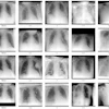
| FIGURE 1.1.1 Sessile tubular adenomas. 3D endoluminal CTC image (A) shows a sessile 6-mm polyp in the ascending colon, which is confirmed on the transverse 2D CTC image (B, arrow) and was removed at same-day optical colonoscopy (C). 3D endoluminal CTC image (D), coronal 2D CTC image (E), and digital photograph from corresponding colonoscopy (F) from a different patient show another typical sessile polyp in the sigmoid colon (8-mm tubular adenoma). |
Atlas of Gastrointestinal Imaging Figure 1.1.1 Sessile tubular adenomas.
Latest in Home

Musculoskeletal strain affects majority of radiologists
April 23, 2026

Sponsored
Turning Innovation into Everyday Efficiency
April 22, 2026
GAE benefits may just be ‘placebo effect’
April 22, 2026


