Dear Digital X-Ray Insider,
Chest x-rays are usually initially performed to screen for acute thoracic aortic dissection, a sudden life-threatening condition in which blood leaks from a tear in the aorta.
Yet the diagnostic accuracy of this method is low, and CT is the established superior modality for this indication.
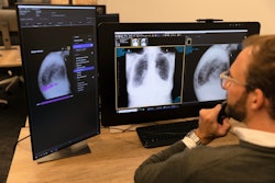
Could an artificial intelligence (AI) algorithm improve the performance of chest x-ray in cases of acute thoracic aortic dissection? Researchers in South Korea believe so, and they recently argued that such an algorithm could have a significant clinical impact, given that x-ray is an easily accessible diagnostic tool in emergency settings. Check out the story on the research in this edition's Insider Exclusive.
Speaking of AI, it led the way in digital x-ray research presented at RSNA 2022, and we were on hand in Chicago to highlight the news. One of the most read articles we published covered fundamental considerations stakeholders need to consider before integrating AI tools into the clinical workflow.
Below are a few other studies we covered at RSNA 2022 in which x-ray imaging took center stage:
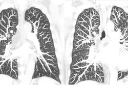
- Dark-field chest x-ray and conventional x-ray went head-to-head for the first time, with dark-field chest x-ray proving more effective for diagnosing pulmonary emphysema.
- A deep-learning model appears to be able to predict 10-year risk of death from heart attack or stroke by using a single chest x-ray.
- A commercially available AI algorithm approved for detecting bone fractures showed high sensitivity in osteoporosis patients with suspected vertebral fractures.
- Research from South Korea shows that AI-based software may improve the detection of lung nodules in countries using x-ray as part of population health check-ups.
Still, while AI continues to be a hot topic, implementing algorithms in clinical practice is slow, with a recent survey from Bournemouth University in England suggesting the jury is still out on how confident radiographers are in using the technology. Meanwhile, a recent study we covered questioned the reliability of bone age AI.
Also, diagnosing COVID-19 on x-ray remains a major area of research, with new techniques under development. A Danish team recently updated a chest x-ray imaging score that could be a valuable tool for measuring the severity of COVID-19 pneumonia.
Finally, with children caught in the crosshairs of the war in Ukraine, experts at Imperial College London's Centre for Blast Injury Studies recently recommended the use of CT over x-ray for identifying significant thoracic injury in children exposed to explosive blasts.
That's all for now. Be sure to check back often for more news in your Digital X-Ray Community.


