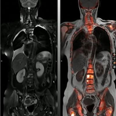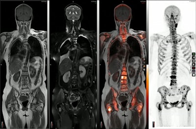
TORONTO - Hybrid PET/MRI imaging could eventually serve as a "one-stop shop" for imaging cancer patients who typically need several imaging studies before surgery, according to research presented at the International Society for Magnetic Resonance in Medicine (ISMRM) annual meeting.
Hersh Chandarana, MD, of New York University's Langone Medical Center in New York City discussed use of the hybrid imaging approach at his hospital during a June 3 session on molecular imaging. The approach has shown promise in a number of abdominopelvic malignancies, such as lymphoma, colorectal malignancy, gynecologic malignancies, and prostate cancer, he said.
"PET/MRI in a clinical practice can be used as a 'one-stop shop' for an oncologic imaging for cancer patients," he said.
Both PET and MRI scanners are used in cancer imaging, with PET/MRI hybrid scanners developed about 12 years ago. These scanners can provide almost simultaneous detailed tumor assessments from multiparametric MRI with complementary physiologic information from PET. Thus, when images are fused -- essentially overlaid on one another -- by software during processing, the approach provides diagnostic value for clinicians not possible with either approach alone. Several studies support the role of PET/MRI for cancer evaluation, he noted.
To illustrate the value of PET/MRI, Chandarana presented a case of a woman with cervical cancer. Initially, she underwent an MRI exam that suggested the cancer had spread beyond her cervix. A CT scan was also performed, with inconclusive results. Ultimately, a PET/MRI was performed, which provided the details necessary for surgery staging.
The example illustrates the concept of the "one-stop-shop" approach -- with a single PET/MRI session (which also can be done quicker and with lower radiation doses, Chandarana noted) potentially replacing the two separate imaging exams in these cases, he said.
However, with hybrid PET/CT scanners also showing great promise, in some instances the question now is which of these hybrid approaches -- PET/MRI or PET/CT -- should be used, Chandarana said.
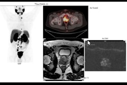
 An image presented during a molecular imaging session at the ISMRM meeting in Toronto showing the range of information a hybrid PET/MRI approach provides. Image courtesy of Hersh Chandarana, MD.
There's no definitive answer to this question, but Chandarana noted a small study comparing the two in cancer patients imaged by both methods on the same day. Findings were noted for PET/CT but not for PET/MRI in just two out of 134 patients, while PET/MRI positively affected clinical management in 24 patients, he said.
Another common question is whether PET provides the same data in a hybrid PER/MRI scanner as it does in a hybrid PET/CT scanner, Chandarana said. On this, he said studies have found that maximum standardized uptake values (SUVmax) -- a standard quantitative PET measurement -- used in PET/MRI studies strongly correlate with SUVmax measured with PET/CT.
"PET from PET/MRI can provide information similar to [PET from] PET/CT," he said.
Ultimately, the "holy grail" is to develop algorithms to provide simultaneous quantitative PET/MRI data that can be used to better characterize tumors not only for tumor surgery staging but also perhaps to help guide and monitor therapies for individualized patient care, Chandarana said.
"We can take advantage of the superior soft tissue contrast that MRI provides over CT for staging tumors and then we can think about how we can combine MRI and PET to get more information from PET to better understand tumor biology," he concluded.


