Researchers from Washington University in St. Louis have developed a new imaging technique based on optical tomography (OCT) combined with deep learning that may provide accurate, real-time, computer-aided diagnosis of colorectal cancer.
In the pilot study, to be published in Theranostics, Quing Zhu, a professor of biomedical engineering in the McKelvey School of Engineering, and Yifeng Zeng, a biomedical engineering doctoral student, used the technique on more than 26,000 individual frames of imaging data from colorectal tissue samples. Compared with pathology reports, they were able to identify tumors with 100% accuracy, according to a Washington University statement.
OCT detects differences in the way healthy and diseased tissue refract light, and it's highly sensitive to precancerous and early cancer morphological changes, the researchers noted. When further developed, the new technique could be used as a real-time, noninvasive imaging tool alongside traditional colonoscopy.
When surgeons use colonoscopy to examine the colon surface, this technology could be zoomed in locally to help make a more accurate diagnosis of deeper precancerous polyps and early-stage cancers versus normal tissue, according to senior author Zhu.
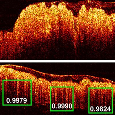
The researchers used RetinaNet, a neural network model of the brain where neurons are connected in complex patterns to process data to recognize and learn the patterns in tissue samples. They trained and tested the network using about 26,000 OCT images acquired from 20 tumor areas -- 16 benign areas and six abnormal areas in patient tissue samples. They found benign tissue displayed a teeth-like pattern. The diagnoses predicted by this system were compared with evaluation of the tissue specimens using standard histology, resulting in a sensitivity of 100% and a specificity of 99.7%.
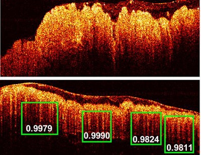 The OCT imaging detected images of colon cancer (top photo) and normal colon tissue. The green boxes indicate the scores of probability of the predicted "teeth" patterns in the tissue. Image courtesy of Zhu Lab.
The research team is now developing a catheter that could be used simultaneously with the colonoscopy endoscope to analyze the teeth-like pattern on the surface of the colon tissue and provide a score of the probability of cancer from RetinaNet to surgeons.


