Dear Artificial Intelligence Insider,
The challenge in assembling image datasets large enough to train artificial intelligence (AI) algorithms for specific use cases has inspired researchers and software developers to explore a variety of different training methods.
In one such approach, researchers from Stanford University have found that pretraining a 3D convolutional neural network with a dataset of over 500,000 annotated YouTube clips of natural videos significantly improved the performance of their model for detecting appendicitis on CT exams.
Their technique could also potentially generalize to other imaging applications for cross-sectional imaging modalities, according to this edition's Insider Exclusive.
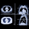
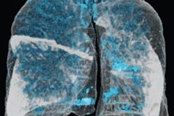
In other news, an AI algorithm is showing potential as a tool for image-based diagnosis and quantification of emphysema severity. A multinational team reported that a prototype algorithm for unenhanced chest CT exams provided results that correlated strongly with pulmonary function tests.
When it comes to screening mammography, the combination of multiple AI algorithms and a radiologist yields the best results, according to a large study. Other researchers concluded, however, that AI could not only outperform radiologists but also improve their performance.
New research has also shown that AI-based assessment of body composition biomarkers on abdominal CT scans could be used to more accurately stratify risk for future serious adverse events or death in asymptomatic patients than traditional parameters, offering promise for further augmenting the value of CT scans. After quantifying blood flow on cardiac MRI, an AI algorithm can also predict -- perhaps better than a physician can -- if a patient will go on to have a heart attack or stroke.
The U.S. Food and Drug Administration (FDA's) clearance of Caption Health's AI-based echocardiography acquisition application created a new regulatory classification for AI-based image acquisition and optimization software. An FDA representative detailed the new rule covering this new CAD device type -- computer-aided acquisition/optimization (CADa/o) -- in a talk during a public workshop on the evolving role of AI in radiology. In another presentation during the FDA's two-day workshop, Dr. Peter Chang of the University of California, Irvine discussed the key issues that are limiting AI's current clinical utility in radiology.
What do patients think about the use of AI in radiology? They're cautiously optimistic, Canadian researchers reported. AI can also improve reader performance on automated breast ultrasound exams and predict progression of osteoarthritis on x-rays.
In addition, AI can aid in the management of stroke patients. For example, a machine-learning algorithm can combine with radiomics to determine on CT if an ischemic stroke in the basal ganglia occurred within the time window during which a stroke patient can still receive thrombolysis. AI-based analysis of multiparametric MRI exams can also find out if a stroke patient's symptoms began within the thrombolysis treatment window.
Business analytics based on AI can create value across the imaging value chain in 14 ways, according to Dr. Teresa Martin-Carreras of the Hospital of the University of Pennsylvania in Philadelphia and Dr. Po-Hao Chen of the Cleveland Clinic.
Do you have an idea for a story you'd like to see covered in the Artificial Intelligence Community? Please feel free to drop me a line.


