Dear AuntMinnie Member,
It's like something from the holodeck on Star Trek: The Next Generation: A walk-in virtual reality room that features massive representations of human anatomy, giving physicians the ability to interact with medical images in ways never before imagined.
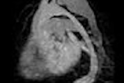
Is this the product of some Hollywood scriptwriter's overactive imagination? Nope, it's a real working prototype in operation at a Dutch hospital. Researchers at the facility, Erasmus Hospital in Rotterdam, are exploring the system's potential for 3D echocardiography, according to an article by staff writer Eric Barnes in our Cardiac Imaging Digital Community.
The group wanted to see if the 3D viewing room could solve what some perceive to be a problem in 3D advanced visualization -- 3D images that are displayed in 2D formats, either on flat monitors or film printouts. Could viewing 3D cardiac images in a virtual reality room reveal additional information not found with more conventional display modes? Find out what they discovered by clicking here.
In other news in the community, we present the work of researchers from Mount Sinai School of Medicine in New York City, where a group has been investigating the use of PET/CT with FDG to analyze the composition of plaques in the carotid arteries. Other studies have found that the type of plaque is often more important than degree of arterial stenosis in determining a patient's risk of a future cardiac event.
PET/CT's combination of anatomic and functional data helped them develop some interesting findings on the relationship between plaque composition and cardiac risk. Learn more from our article by staff writer Wayne Forrest by clicking here.
Get these stories and more in the Cardiac Imaging Digital Community, at cardiac.auntminnie.com.


