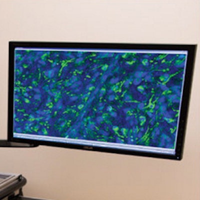
A new microscopy technique sorts healthy from malignant brain tumors, making surgery safer and more precise, according to the U.S. National Institute of Biomedical Imaging and Bioengineering (NIBIB).
Surgeons want to remove as much tumor as possible during brain surgery, and to do so, they can ask pathologists to examine tissue samples during surgery to inform decisions. But even assuming a pathologist is available, prepping a sample takes more than a half hour, increasing costs and risks to the patient, NIBIB said in a statement.
The new technique not only speeds up the tissue-analysis process, it also enables automatic processing to be performed remotely. It cuts processing time and could significantly increase the accuracy of brain tumor surgery in the operating room, according to Behrouz Shabestari, PhD, director of the NIBIB program in optical imaging and spectroscopy.
"It basically optimizes the surgical result and has the potential to improve patient outcomes by increasing safety and survival rates," he added.
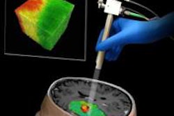
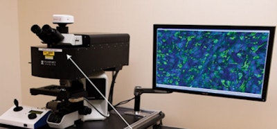 A stimulated Raman scattering (SRS) microscope helps neurosurgeons analyze tissue without the need for tissue processing, speeding up the decision-making process during brain tumor surgery. Image courtesy of NIBIB.
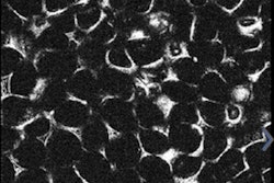
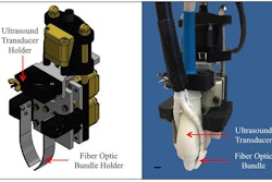
Lead author Dr. Daniel Orringer and colleagues from the University of Michigan used a microscopy technique called stimulated Raman scattering (SRS) that eliminates the need for tissue processing, or slicing and staining the tissue. By using a fiber-laser microscope similar to the fiber optics in internet and phone cables, as well as developing a way to reduce background signals common to fiber-laser images, the group created a high-resolution system that has been validated in more than 100 patients.
The researchers also developed another technique called stimulated Raman histology (SRH). SRH is used to process the resulting images quickly by taking advantage of the chemical properties of the tissue, making proteins and DNA appear purple and lipids appear pink.
The result is similar to using hematoxylin and eosin (H&E) staining, and pathologists can interpret the images without special training. Eliminating sectioning and staining reduces processing time to about three minutes, 10 times faster than standard tissue-processing techniques. In a 30-sample analysis, pathologists came to similar conclusions using SRH and conventional techniques.


