CHICAGO - Digital x-ray and PACS vendor Fujifilm Medical Systems USA of Stamford, CT, is entering the market for mobile imaging with FCR Go, a portable system that features a Carbon XL computed radiography (CR) reader and a notebook version of the company's Flash IIP technologist console, integrated into a mobile cart supplied by Hitachi of Japan.
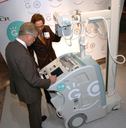 |
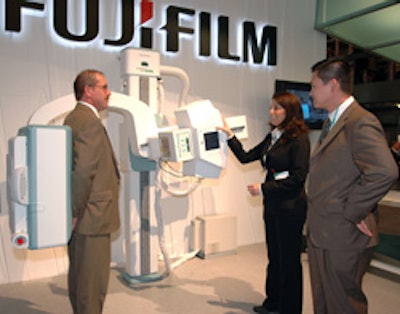
| Fuji is highlighting FCR Go in its RSNA booth. |
Fuji is also highlighting the system's impact on technologist workflow. The integrated IIP console enables RTs to look at images remotely and order retakes if necessary. In addition, the system enables radiologists who are already reading images produced on stationary CR systems to enjoy the same look and feel for their mobile images.
FCR Go has a throughput of 94 imaging plates per hour and a 15-kW x-ray generator, and preview images can be produced in 23 seconds. The system also supports the DICOM Worklist protocol. Fuji is showing the unit as a work-in-progress, with availability expected in mid-2008. Hitachi is already selling FCR Go in Japan.
Carbon XL-2 is a new version of the company's Carbon XL reader, with the major new feature being 50-micron resolution for 18 x 24-cm and 24 x 30-cm imaging plate sizes. The company believes the reader will be well-suited for clinical applications that require high-resolution imaging, such as extremity work or orthopedics.
On the software side, Fuji is touting version 5.0 of its Flash IIP console software. The new version enables the output of 12-bit CR and DR images to PACS displays that support the larger data formats. The new software also includes customized values of interest lookup tables (VOI LUTs) and log linear amplification.
The new capabilities give radiologists better image quality and improved diagnostic confidence as they ensure enhanced visualization in all areas of the gradation curve at PACS workstations, according to the company. Version 5.0 also allows Fuji's digital x-ray systems to address PACS and networks of different sophistication levels by providing scalable options for file size and bit depth, the company said.
Fuji began shipping its FCRm system for full-field digital mammography (FFDM) applications in the U.S. in 2006, and more than 4,000 systems have been shipped globally. At this year's RSNA meeting, Fuji is highlighting the comparable image quality between its CR-based FFDM systems and flat-panel DR mammography.
The company is also showing a new version of its Synapse mammography workstation that includes advanced tools for mammography image review, such as a "mirror image" mode for current/prior image review that transfers changes made on one image to the corresponding image on the display.
As a work-in-progress, Fuji is discussing lossy compression of mammography images at ratios in excess of 20:1, which could help foster telemammography by reducing image transfer time and archiving costs. The company has a study under way at Elizabeth Wende Breast Clinic in Rochester, NY, to determine if clinicians find the compressed images acceptable for diagnostic use. Fuji is also showing its CR mammography images displayed on a 16-megapixel monitor sourced from Planar Systems of Waltham, MA.
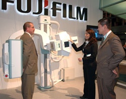 |
| Fuji's Unity SpeedSuite DR system. |
PACS
In PACS, Fuji is introducing the latest version of its Synapse software, version 3.2.1. The software is an upgrade of version 3.2 and offers enhancements to increase user efficiency and productivity, provide greater functionality for interpreting digital mammography studies, and integrate with cardiology, 3D imaging, and PET/CT.
Synapse 3.2.1's new graphical user interface (GUI) is designed to help radiologists more quickly identify and access information and generate reports. The interface's new look is based on Fuji’s Web-based technology for enhanced ease of use for all clinical users, including mammographers and cardiologists. Specific GUI changes include increased use of color to help identify changes in information on the display, new icons, and a contrast scheme that is suitable for radiology viewing conditions.
Other enhancements are designed for FFDM, such as quadrant zooming and common frame of reference (CFOR) navigation, which allows for window leveling, zooming, and mirror panning of linked images to improve diagnostic efficiency.
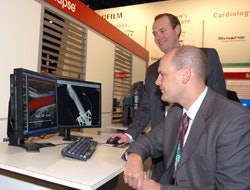 |
| Fuji is featuring the new version of its Synapse PACS software. |
Fuji is also showing advanced cardiology capabilities from its January 2007 acquisition of ProSolv CardioVascular. The Synapse ProSolv CardioVascular application, scheduled for release in the first quarter of 2008, enables radiologists to access all cardiology images and information while working within Synapse. Cardiologists also can access all Synapse radiology data from one workstation with a single sign-on and familiar user interface.
The Synapse ProSolv CardioVascular application is available with ProSolv's version 4.0, which is being shown as a work-in-progress and is scheduled for release in the first quarter of 2008. Features on version 4.0 will include viewing and reporting enhancements for cardiac catheterization and echocardiography.
Fujifilm also is demonstrating future extensions to its distributed 3D software, including PET/CT fusion. Work-in-progress advancements to existing diagnostic maximum intensity projection and multiplanar reformatting (MIP/MPR) capabilities for Synapse are on display as well.
By Brian Casey and Wayne Forrest
AuntMinnie.com staff writers
November 27, 2007
Related Reading
Fuji launches mobile CR package, November 13, 2007
Road to RSNA, PACS Accessories, Fujifilm Medical Systems USA, October 30, 2007
Road to RSNA, Women's Imaging, Fujifilm Medical Systems USA, October 26, 2007
Road to RSNA, PACS, Fujifilm Medical Systems USA, October 25, 2007
Fuji wins Premier DR contract, October 11, 2007
Copyright © 2007 AuntMinnie.com
