Myocarditis:
Clinical:
Myocardial inflammation (myocarditis) can be secondary to a number of etiologies including infection (typically viral or HIV), toxic agents (drugs, chemotherapy), or post-transplant rejection. For viral myocarditis, spontaneous recovery within a few weeks to months is common [3]. However, chronic myocarditis (dilated cardiomyopathy) occurs in up to 21% of cases [3].
X-ray:
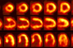
MR: With acute myocarditis, delayed hyperenhancement is generally nodular and subepicardial, without respect for vascular territories, and often with an inferolateral/lateral and apical distribution [1,2]. The changes may be subtle and focal acutely, becoming more diffuse over the next 10 days [1]. In myocarditis, myocardial damage is more diffuse than damage due to infarction. Islands of necrotic cells are scattered throughout the focus of acute myocarditis [2]. Therefore, contrast enhancement in acute myocarditis may not be as intense as in myocardial infarction [2]. During healing, inflammatory cells infiltrate the myocardial regions of myocarditis, and eventually necrotic myocytes are replaced by areas of fibrous tissue and delayed contrast enhancement can persist in the chronic phase [2]. The area of myocarditis diminishes in size as it is replaced by scar and this can explain the observation that contrast enhancement typically decreases over time [2].
REFERENCES:
(1) AJR 2007; Lim RP, et al. Non-ischemic causes of delayed myocardial hyperenhancement on MRI. 188: 1675-1681
(2) Radiographics 2006; Vogel-Claussen J, et al. Delayed enhancement MR imaging: utility in myocardial assessement. 26: 795-810
(3) Radiology 2008; Gutberlet M, et al. Suspected chronic myocarditis at cardiac MR: diagnostic accuracy and association with immunohistologically detected inflammation and viral persistence. 246: 401-409
