Radiol Clin North Am 1996 Jan;34(1):97-117. Pulmonary complications after bone marrow transplantation.
Department of Diagnostic Imaging, St. Jude Children's Research Hospital, Memphis, Tennessee, USA.
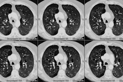
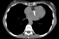
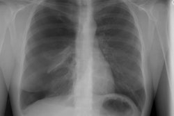
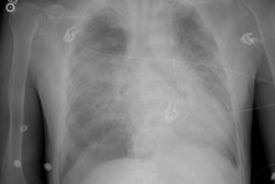
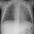
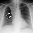
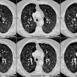
Many of the pulmonary complications that we have described have a nonspecific radiographic appearance. The most crucial information for proper interpretation of the chest radiographs is the chronologic onset of radiographic abnormalities after transplantation. Before and immediately after engraftment, local peripheral opacities accompanied by a surrounding rim of edema are regarded as fungal infections, and therapy with granulocyte transfusions and amphotericin B is initiated. Diffuse interstitial thickening is likely to represent edema, pulmonary hemorrhage, bacterial infection, or ARDS rather than CMV or P. carinii pneumonia in the neutropenic host. After engraftment, diffuse interstitial processes become the predominant lung abnormalities. In allogeneic transplant patients who are serologically positive for CMV or who receive serologically positive donor marrow for CMV, pneumonitis caused by this virus is perhaps the most common treatable lung infection. Idiopathic interstitial pneumonias present in a similar fashion to CMV pneumonia; however, the response to corticosteroid therapy is only occasionally gratifying. The onset of nodular opacities in this period may be due to a number of disorders, such as opportunistic infection, BOOP, PTLPD or recurrent tumor. Open lung biopsy usually is required for definitive diagnosis.
PMID: 8539356, MUID: 96133839
