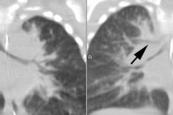
Bronchogenic Cyst:
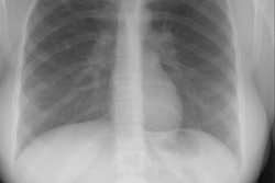
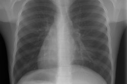
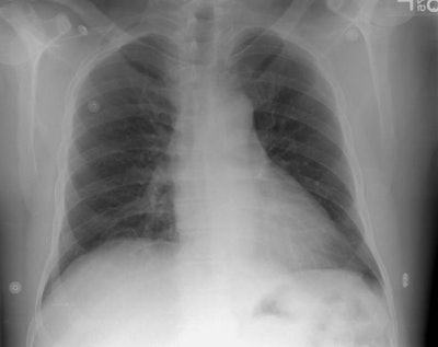
The patient shown in the case below is a 55 year old man who presented with left sided pleuritic chest pain. The radiograph demonstrated a rounded density over the medial aspect of the left hemidiaphragm (not well seen on the lateral view). A previous chest radiograph 4 months earlier did not demonstrate this abnormality. The patient had no signs of infection, and a smoking history. A CT of the chest was performed to evaluate this finding.
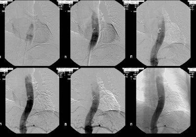
The CT scan demonstrated a low density mass in the left posterior costophrenic sulcus. A percutaneous biopsy was performed and on aspiration material with the consistency of "crank oil" was withdrawn. The fluid contained inflammatory cells, but no organisms. Surgical removal was recommended. Because of the possibility that the lesion may represent a bronchopulmonary sequestration, a pulmonary arteriogram was also performed.
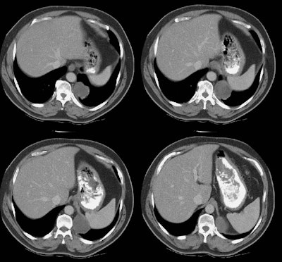
The exam demonstrated no systemic arterial supply to the lesion. At histopathologic analysis the lesion was found to be a bronchogenic cyst.
