| J Nucl Med 1995 Apr;36(4):581-585 |
Posterolateral defect of the normal human heart investigated with
nitrogen-13-ammonia and dynamic PET.
de Jong RM, Blanksma PK, Willemsen AT, Anthonio RL, Meeder JG, Pruim J, Vaalburg W, Lie
KI.
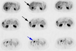
The posterolateral defect is a common artifact seen when static 13N-ammonia imaging with
PET is used to assess myocardial perfusion. The aim of this study was to compare dynamic
and static. 13N-ammonia PET and to obtain more insight into the cause of the
posterolateral defect. METHODS: Dynamic 13N-ammonia PET was performed in 19 healthy
nonsmoking volunteers at rest. Perfusion was assessed in the early phase of the study
using a curve fit method over the first 90 sec. Nitrogen-13 accumulation (static PET) was
assessed 4 to 8 min after injection. Each study was normalized to a mean of 100. The
average distribution of normalized perfusion and activity was calculated in 24 segments.
Heterogeneity of both activity and perfusion distribution were assessed and the activity
distribution was compared with perfusion distribution. RESULTS: Perfusion distribution was
homogeneous, with the exception of the inferior and apical regions. Activity distribution
was inhomogeneous, with a lower activity in the posterolateral and apical regions. In the
whole left ventricle, significant differences in distribution were found between static
and dynamic imaging. CONCLUSION: Perfusion distribution was significantly different on
dynamic images compared to static images. The posterolateral defect was not found on
dynamic images. The posterolateral defect and other inhomogeneities in activity
distribution are caused by tracer-dependent features, probably a redistribution of
metabolites of 13N-ammonia.
