Respiratory viral infections in lung transplant recipients: radiologic findings with clinical correlation.
Matar LD, McAdams HP, Palmer SM, Howell DN, Henshaw NG, Davis RD, Tapson VF
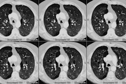
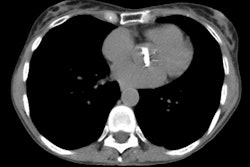
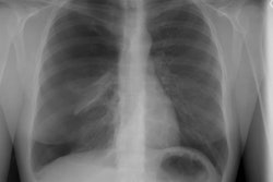
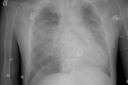
PURPOSE: To evaluate radiologic finding of respiratory viral infection in lung transplant recipients with clinical correlation. MATERIALS AND METHODS: Over 5 years, 21 episodes of respiratory viral infection (parainfluenza [n = 9], respiratory syncytial virus [n = 8], adenovirus [n = 5], influenza [n = 2]) were diagnosed 6-727 days (mean, 270 days) after lung transplantation in 20 recipients. Chest radiographs, computed tomographic (CT) images, and clinical records were reviewed. RESULTS: Sixteen episodes of respiratory viral infection were diagnosed in patients with symptoms of lower respiratory tract infection or acute allograft dysfunction; five were diagnosed in asymptomatic patients. Chest radiographs were abnormal in 11 (52%) episodes; findings included heterogeneous or homogeneous opacities and masslike consolidation. All patients with radiographic abnormalities were symptomatic. Chest radiographs were unchanged from baseline in 10 (48%) episodes; in one, CT revealed findings not depicted at radiography. Adenoviral infection (n = 5) was typically symptomatic, was associated with new radiographic abnormalities, and was rapidly lethal (n = 4). Infection with parainfluenza and/or respiratory syncytial virus was commonly asymptomatic and was not associated with radiographic abnormalities; affected patients had good outcomes. CONCLUSION: Respiratory viral infections are important causes of morbidity and mortality in lung transplant recipients. Radiographic abnormalities in patients with respiratory viral infections were usually accompanied by symptoms of lower respiratory tract infection. Adenoviral infection was frequently accompanied by progressive pulmonary opacity and fatal outcome.
