Preoperative and postoperative imaging in the surgical management of pulmonary emphysema.
Slone RM, Gierada DS, Yusen RD
For patients with emphysema, imaging studies have been useful for diagnostic
purposes and for preoperative patient selection for surgical intervention,
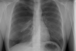
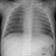
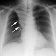
such as bullectomy, lung transplantation, and LVRS. Chest radiography is
useful in evaluating hyperinflation. Inspiratory and expiratory films are
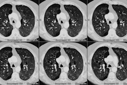
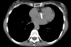
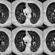
used to estimate diaphragmatic excursion and air-trapping. CT scan is used
to evaluate the anatomy and distribution of emphysema throughout the lungs,
providing information clinically unobtainable by other means. Both imaging
techniques are useful for detecting other disease processes. Radionuclide
lung scanning also provides an estimate of target areas, volume occupying
but nonfunctioning lung. Cohort studies utilizing these imaging techniques
have demonstrated associations between preoperative characteristics and
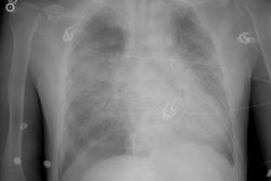
postoperative outcome. The imaging studies, especially the chest radiograph,
have also played an important role in postoperative management. Many other
imaging options are available, such as HRCT scan, quantitative CT scan,
and single photon emission CT scan. Other techniques, such as MR imaging,
may play a future role as well.
Publication Types:
Review
Review, tutorial
