Bronchiectasis: accuracy of high-resolution CT in the differentiation of specific diseases.
Cartier Y, Kavanagh PV, Johkoh T, Mason AC, Muller NL
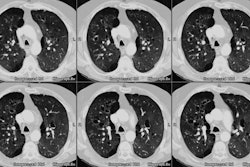
OBJECTIVE: The aim of the study was to determine whether various causes of bronchiectasis can be differentiated by the pattern and distribution of abnormalities seen on high-resolution CT. MATERIALS AND METHODS: The retrospective study included 82 consecutive patients who had a specific diagnosis of bronchiectasis proven by appropriate clinical and laboratory criteria. All patients underwent high-resolution CT scanning (1- to 1.5-mm collimation). The CT scans were assessed for the presence, extent, type, and anatomic distribution of bronchiectasis by two independent observers who were not aware of the clinical data. The observers recorded their most likely diagnosis and the degree of confidence in that diagnosis. RESULTS: The two independent observers made a correct diagnosis in 61% of cases (100/164 interpretations). On average, a correct diagnosis was made in 19 (68%) of 28 cases of cystic fibrosis, 16 (67%) of 24 cases of previous tuberculosis, six (43%) of 14 cases of previous childhood infection, five (56%) of nine cases of allergic bronchopulmonary aspergillosis, and four (57%) of seven cases of other causes of bronchiectasis. We found moderate agreement between the observers for the correct diagnosis (kappa = .53) and good agreement for the presence or absence of bronchiectasis in each lobe (kappa = .71). CONCLUSION: The pattern and distribution of abnormalities revealed by high-resolution CT in patients with bronchiectasis are influenced by the underlying cause. Bilateral, predominantly upper lobe, bronchiectasis is seen most commonly in patients with cystic fibrosis and allergic bronchopulmonary aspergillosis, unilateral upper lobe predominance in patients with tuberculosis, and lower lobe predominance in patients after childhood viral infection.
