Radiographics 2000 May-Jun;20(3):713-23
Imaging features of primary and recurrent esophageal cancer at FDG PET.
Skehan SJ, Brown AL, Thompson M, Young JE, Coates G, Nahmias C.
Because of the poor prognosis for patients with esophageal cancer and the risks associated
with surgical intervention, accurate staging is essential for optimal treatment planning.
Positron emission tomography (PET) with 2-[fluorine-18]fluoro-2-deoxy-d-glucose (FDG) is a
useful adjunct to more conventional imaging modalities in this setting. FDG PET is not an
appropriate first-line diagnostic procedure in the detection of esophageal cancer and is
not helpful in detecting local invasion by the primary tumor, and further studies are
required to determine its efficacy in the detection of local nodal metastases. However,
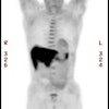
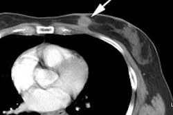
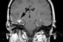
FDG PET is superior to anatomic imaging modalities in the ability to detect distant
metastases. Metastases to the liver, lungs, and skeleton can readily be identified at FDG
PET. In addition, FDG PET has proved valuable in determining the resectability of disease
and allows scanning of a larger volume than is possible with computed tomography.
Recurrent disease is readily diagnosed and differentiated from scar tissue with FDG PET.
In addition, FDG PET may play a valuable role in the follow-up of patients who undergo
chemotherapy and radiation therapy, allowing early changes in treatment for unresponsive
tumors. The management of most patients with esophageal cancer can be improved with use of
FDG PET.


