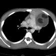
Lymphangioma:
Clinical:
Most lymphangiomas are present at birth and are usually detected within the first 2 years of life. They usually occur in the head, neck, and axilla. Approximately 10% of cervical lymphangiomas extend into the mediastinum. Fewer than 1% of lymphangiomas are limited only to the mediastinum.Primary mediastinal lymphangioma is rare usually presents as a solitary mediastinal mass. The lesion is most frequently seen in men. Because the lesion grows slowly and has a soft consistency, patients are usually asymptomatic. The majority of lesions are cystic and are located in the superior mediastinum adjacent to the right lateral wall of the trachea. Because of their insinuating nature, complete surgical resection may be difficult [2]. Depending on the lesions location differential considerations include a bronchogenic cyst, a foregut duplication cyst, pericardial cyst, thoracic duct cyst, thymic cyst, or cystic teratoma.
Histologically lymphangiomas are divided into three groups depending on the size of the lymphatic channels. Capillary (simple) lymphangiomas are composed of small lymphatics; cavernous lymphangiomas of larger lymphantics; and cystic lymphangiomas of large, macroscopic lymphatic spaces. The tumors are usually thin-walled, multilocular, lined by endothelial cells, and contain clear yellow fluid.
X-ray:
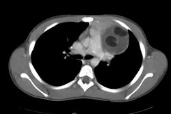
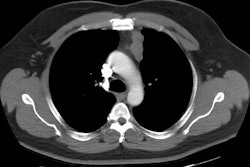
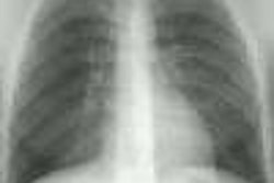
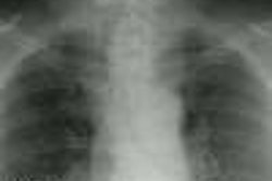
On CT the lesion classically appears as a well circumscribed, low density lesion molding to the mediastinal contours and enveloping the great vessels. The most common finding, however, is a rounded, low density mass adjacent to the right lateral wall of the trachea. Increased density or inhomogeneity may be seen secondary to internal hemorrhage. The lesions generally do not enhance after IV contrast administration. Calcification is very rare and the tumor characteristically does not displace the mediastinal structures. On MR imaging, the lesions usually has heterogeneous signla on T1 images, and high signal on T2 images [2].REFERENCES:
(1) J Thorac Imag 1996;11 83-85
(2) Radiographics 2002; Jeung MY, et al. Imaging of cystic masses of the mediastinum. 22: S79-S93
