Mesothelioma:
Clinical:
Diffuse malignant mesothelioma is the most common primary pleural
neoplasm (metastatic lesions are the most common pleural
malignancy overall). Even though mesothelioma accounts for over
90% of primary pleural malignancies [14], it is still a rare
lesion. The tumor affects an estimated 2,500 Americans annually
[10]. Approximately 80% of mesotheliomas arise from the parietal
pleura and on histologic analysis the cell type is often mixed and
difficult to distinguish from adenocarcinoma. Some mesotheliomas
(about 20%) contain hyaluronic acid which will stain with
colloidal iron and help to differentiate the lesion from an
adenocarcinoma. Positive vimentin staining in the fibrous
component of the tumor, the absence of mucin, and the absence of
carcinoembryonic antigen also suggest mesothelioma.There are three
main histologic types of mesothelioma: epitheliod (55-60% of
cases), sarcomatoid (10-15%), and biphasic or mixed (20-35%) [14].
The nonepitheliod types have a worse prognosis [14].
Risk Factors:
There is an increased risk for mesothelioma associated with asbestos exposure. Up to 80% of cases of mesothelioma occur in patients with a history of asbestos exposure (short or long term) and the latent period is about 20 to 40 years (about 20% of patients that develop mesothelioma do not have any relevant asbestos exposure history [14]). The risk has been shown to increase with the duration and intensity of exposure [15]. The lifetime risk/incidence for developing mesothelioma in heavily exposed individuals in as high as 10% [12,15]. Asbestos fibers can be subdivided into two large groups- amphiboles (stiff and straight) and serpantines (curly and flexible) [15]. The carcinogenicity of asbestos fibers is proportional to the fibers aspect ratio (the ratio of the fiber length to width), with higher aspect ratios confering higher tumorigenicity [15]. The amphiboles asbestos fiber "crocidolite" has the greatest association with eliciting mesothelioma and bronchogenic carcinoma. The serpentine fiber "Chrysotile" accounts for approximately 80% of the asbestos used in the Western World- it tends to be deposited in the proximal airways, is relatively water soluble, and has a strong association with the formation of pleural plaques. It is postulated that exposure to pure chrysotile does not cause malignant mesothelioma, however, contaminants such as tremolite (an amphibole fiber) may be responsible for tumor development in these individuals. There is no recognized threshold below which malignant mesothelioma will not occur, however, there is no proof that low concentrations of asbestos will cause mesothelioma. Greater intensity and duration of exposure are associated with a higher incidence of malignancy [15]. Cigarette smoking does not affect an asbestos exposed individuals risk for mesothelioma. The actual process by which the asbestos fiber incites malignancy is poorly understood.
Other associations with mesothelioma include chronic inflammation
and scarring as in cases of tuberculosis and empyema, and possibly
radiation exposure in patients who have a history of XRT for
Hodgkin's disease.Simian virus 40 (an oncogenic DNA virus) has a
controversial association [15].
Physical Exam:
Mesothelioma most commonly occurs in patients between 50-70 years
and is more common in men than women (4:1) [15]. Clinically
patients present with the insidious onset of dyspnea (50%), chest
wall pain (60% and almost always non-pleuritic), cough, and weight
loss. Paraneoplastic syndromes associated with the lesion are rare
but include intermittent hypoglycemia (fewer than 10% of
patients), hypertrophic osteoarthropathy (fewer than 10% of
patients), and clubbing. Thrombocytosis is found in 30-40% of
patients. Neoplastic involvement of the hilar and mediastinal
nodes is present in about 40-45% of patients at autopsy.
Hematogenous metastases to the lung are very uncommon. The pleural
effusion is typically exudative and may be hemorrhagic. A
hyaluronic acid level greater than 0.8 mg/mL is said to establish
the diagnosis of mesothelioma. Serum mesothelin-related protein
(SMRP) is found to be elevated in 84% of patients with malignant
mesothelioma and in less than 2% of patients with other pleural
diseases [12]. A meta-analysis of studies investigating the
association between SMRP and MPM demonstrated a sensitivity of 64%
and a specificity of 89% for diagnosis of MPM [15].
Diagnosis:
Cytologic evaluation of the associated pleural effusion is positive in only about 25% of cases of mesothelioma [6]. Reverse-bevel needle biopsy of the pleura has a sensitivity between 21-43% for the diagnosis of mesothelioma [6]. Percutaneous needle biopsy performed under CT guidance has been reported to have a limited sensitivity (25-60%) in the diagnosis of malignant mesothelioma (usually due to the small sample size), however, sensitivities up to 86% have been reported [11]. Mesothelioma can extend out of the chest cavity through needle biopsy and chest tube tracts [6,9]. Tumor seeding of the needle tract has been reported to occur in about 4-20% of patients [6,11] and preventative local XRT is now used to prevent this occurrence. Using a coaxial biopsy technique for CT directed biopsy can result in a lower incidence of tumor seeding (4%) [11].
Video assisted thoracoscopic surgery (VATS) is the diagnostic procedure of choice (sensitivity 90%). XRT is also performed to all entry ports following the procedure.
Staging / Treatment / Prognosis:
The most widely accepted staging scheme for malignant mesothelioma has been the classification by Butchart et al- however, Stage I disease has not been uniformly associated with an improved prognosis and Boutin has suggested a modification to the Butchart staging because visceral pleural involvement is usually associated with a worse prognosis. A new TNM staging system for mesothelioma has been proposed based upon a more aggressive surgical approach to the lesion [1].
A tumor of any size may be resectable if it is confined to one hemithorax and shows only superficial invasion of the diaphragm or visceral pericardium, localized invasion of the chest wall limited to a previous biopsy site, and a smooth uninvolved inferior diaphragmatic surface. Patients with T1a or T1b lesions are considered potentially curable with surgery. Patients with T2 or T3 lesions may benefit from surgery, but may not be cured [8]. Patients with T4 lesions, N3 nodal disease, or distant metastases are not resectable [9]. The most reliable CT and MR features indicating resectability are a clear fat plane between the inferior surface of the diaphragm and the adjacent abdominal organs and a smooth inferior diaphragmatic contour.
In patients with Stage I disease an attempt to remove all the tumor is performed by an extrapleural pneumonectomy which consists of en block resection of the parietal and mediastinal pleura, lung, ipsilateral pericardium, and the hemidiaphragm. There is a high morbidity and mortality associated with the procedure. Additionally, although disease free survival is improved, no study has demonstrated a significantly improved survival following the procedure. Pleurectomy and decortication is a lung sparing procedure that invovles removal of the parietal and visceral pleura from the lung surface, mediastinum, pericardium diaphragm, and chest wall [15]. The procedure is associated with a higher post surgical recurrence rate than extrapleural pneumonectomy, but metastatic disease is less common [15]. Malignant mesothelioma is relatively radioresistant and response to chemotherapy is poor, however, multimodality therapy following surgery has been shown to prolong survival [9,13]. Pleurectomy (stripping of the pleura and pericardium on the involved side) is usually only a palliative procedure performed in patients with Stage II disease or higher.
Overall prognosis is dismal with an average 9-17 month survival [15] (but up to 28 months in post-surgical Stage IA patients). Newer chemotherapy agents such as pemetrexed, gemcitabine, and vinorelbine have been shown to improve symptoms and prolong survival [10]. Factors associated with a reduced survival time include intrathoracic lymph node metastases, distant metastases, extensive pleural involvement, and age over 75 years [9,15]. Epithelial subtype histology is usually associated with a better prognosis [15]. Metastases to the contralateral lung and pleura occur in about 10% of patients [4].
Butchart Staging for Malignant Mesothelioma:
Stage Findings
I Tumor confined to the ipsilateral pleura, lung, or pericardium
II Tumor invading the chest wall or mediastinal structures, or mets to thoracic
lymph nodes
III Tumor extending through the diaphragm to involve the peritoneum or
extra-thoracic lymph node mets
IV Distant hematogenous mets
Boutin Modification of the Butchart Staging System:
(Subdivides Stage I disease, other stages remain unchanged)
IA Tumor confined to the ipsilateral parietal or diaphragmatic pleura
(no visceral pleural involvement)
IB Tumor confined to the ipsilateral parietal, diaphragmatic, or visceral pleura;
lung; or pericardium
Extra-thoracic Mesothelioma:
Mesotheliomas may also arise from the peritoneum (approximately 20% of cases). These tumors are associated with heavy asbestos exposure and associated pleural plaques/fibrosis is found in 50% of cases. Other sites include the pericardium and tunica vaginalis of the testis. A multicystic peritoneal mesothelioma is a less aggressive lesion with little metastatic potential, but a high incidence of local recurrence [7]. It is more common in young or middle aged females and presents as a multiseptated cystic pelvic mass (ddx: ovarian cystadenoma or carcinoma, lymphangioma, endometriosis, epithelial inclusion cyst, or necrotic leiomyoma) [7]. The lesion is not chemo- or radiation sensitive and the treatment is surgical resection [7].
X-ray:
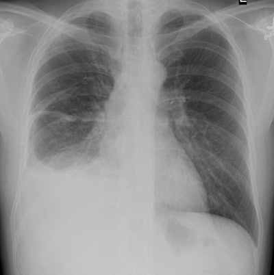
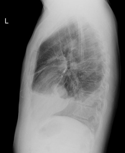
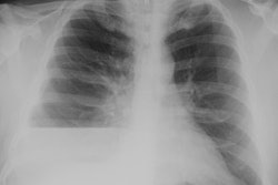
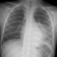
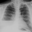
CXR: The lesion usually appears as a markedly thickened, nodular, irregular pleural based mass which coats the pleural surface and grows along fissures. The tumor often encircles the involved lung and is only rarely bilateral. Diffuse pleural thickening can be seen in 60% of cases or pleural masses in 45-60% [15]. Chest wall, diaphragmatic, and mediastinal invasion can be seen with more advanced disease. Contralateral asbestos related pleural disease is seen in 25-50% of cases. A moderate to large, ipsilateral pleural effusion is seen in 50-75% of cases. Because mesothelioma encases the lung it may fix the affected hemithorax so there is no shift away from the side of the tumor, in fact there may be evidence of volume loss on the affected side even in the presence of massive effusion.
|
Mesothelioma on CXR: Middle aged male presented with chest pain and shortness of breath. CXR demonstrated a moderate to large sized right pleural effusion abnormal nodular thickening of the pleura- particularly evident along the fissures. The patient had no known history of asbestos exposure. |
|
|
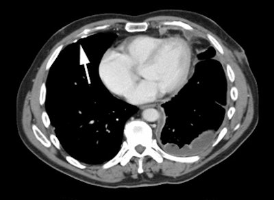
Computed tomography: Common findings include a unilateral pleural effusion, nodular pleural thickening, and interlobar fissure thickening [9]. Circumferential, nodular pleural thickening greater than 1 cm identified in over 90% of cases. Thickening extends into the interlobular fissure in about 85% of cases. Mediastinal pleural involvement is also seen. The absence of pleural thickening does not exclude malignancy, and cases of mesothelioma have been reported in which the only CT finding was a pleural effusion. Calcified pleural plaques are found in about 20% of patients and they may be engulfed by the tumor. Chest wall invasion may manifest as obscured fat planes, invasion of intercostal muscles, or rib destruction. Irregularity of the interface between the chest wall and the tumor is NOT a reliable indicator of chest wall invasion.
|
Mesothelioma on CT: The CT scan on the same patient shown above. The nodular mass-like pleural thickening is readily apparent on CT imaging. |
|
|
Findings suggestive of transdiaphragmatic extension include a soft tissue mass encasing the hemidiaphragm [9], while a clear fat plane between the diaphragm and adjacent abdominal organs and a smooth diaphragmatic contour indicate the tumor is confined to the thorax [9].
Metastases to hilar and mediastinal nodes is present in 40-45% of patients at autopsy [9].
Magnetic resonance imaging: MR provides excellent soft tissue contrast and can aid in evaluation of invasion of adjacent structures/chest wall [12]. On T1 images mesothelioma appears iso- to slightly hyperintense relative to the adjacent chest wall musculature and of moderately increased signal on T2 images [9]. MR is probably superior to CT in determining chest wall, diaphragmatic, or mediastinal invasion, but the staging accuracy is not substantially greater than CT [5,9].
Peritoneal mesothelioma usually produces multifocal bowel obstruction with thickening of the peritoneum/mesentery encasing bowel. Minimal ascites is found (remember, there is often massive ascites associated with carcinomatosis). Mesenteric nodules may be present.
FDG PET: FDG uptake in mesothelioma is significantly greater than in benign pleural disease [8]. Sensitivity has been reported to be 91% and specificity 100%.
REFERENCES:
(2) Radiographics 1996; 16: 613-644
(3) ACR Syllabus #40:p.364
(4) AJR 1998; Montesinos JJ, et al. Thoracic case of the day: Case 2- Malignant pleural mesothelioma. 171: 835-843 (No abstract available)
(5) AJR 1999; Heelan RT, et al. Staging of malignant pleural mesothelioma: Comparison of CT and MR imaging. 172: 1039-1047
(6) Radiology 2001; Adams RF, Gleeson FV. Percutaneous image-guided cutting-needle biopsy of the pleura in the presence of a suspected malignant effusion. 219: 510-514
(7) AJR 2002; Bui-Mansfield LT, et al. Multicystic mesothelioma of the peritoneum. 178: 402
(8) Radiographics 2002; Roach HD, et al. Asbestos: when the dust settles- an imaging review of asbestos-related disease. 22: S167-S184
(9) Radiographics 2004; Wang ZF, et al. Malignant pleural mesothelioma: evaluation with CT, MR imaging, and PET. 24: 105-119
(10) AJR 2006; Armato SG, et al. Variability in mesothelioma tumor response classification. 186: 1000-1006
(11) Radiology 2006; Agarwal PP, et al. Pleural mesothelioma: sensitivity and incidence of needle track seeding after image-guided biopsy versus surgical biopsy. 241: 589-594
(12) Radiographics 2007; Tyszko SM, et al. Best cases from the
AFIP: Malignant mesothelioma. 27: 259-264
(13) AJR 2012; Gill RR, et al. Epithelial malignant pleural
mesothelioma after extrapleural pneumonectomy: stratification of
survival with CT-derived tumor volume. 198: 359-363
(14) AJR 2012; Makis W, et al. Spectrum of malignant pleural and
pericardial disease on FDG PET/CT. 198: 678-685
(15) Radiographics 2014; Nickell LT, et al. Multimodality imaging
for characterization, classification, and staging of malignant
pleural mesothelioma. 34: 1692-1706
