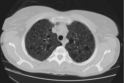
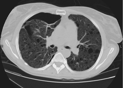
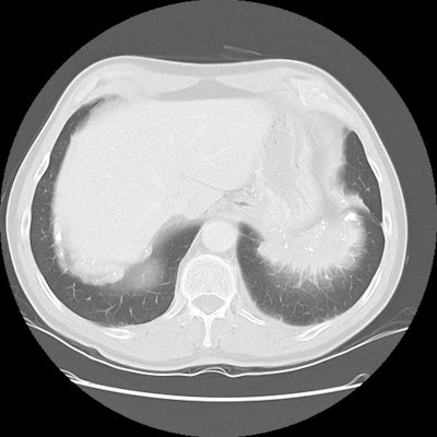
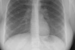
Type I cystic adenomatoid malfomation:
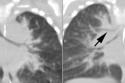
The CT scan below demonstrated a very large cystic lesion occupying the left upper lung. An air-fluid layer can be seen superiorly within the lesion. There is some mediastinal shift to the right and some compressive atelectasis within the adjacent left lung.
