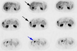
FDG SPECT in patients with lung masses.
Mastin ST, Drane WE, Harman EM, Fenton JJ, Quesenberry L
STUDY OBJECTIVES: To determine whether 2-[fluorine-18]fluoro-2-deoxy-D-glucose (FDG) single-photon emission CT (SPECT) is useful in characterizing pulmonary masses. DESIGN: Scans were prospectively acquired and interpreted. Interpretations were performed with CT or chest radiograph but interpreters were blinded to eventual diagnosis. SETTING: University hospital practice and affiliated Veterans Administration medical center. PATIENTS OR PARTICIPANTS: Forty patients participated as part of an institutional review board-approved research protocol, and informed consent was obtained in all. Eight additional patient scans were acquired as part of their clinical evaluation for pulmonary mass. MEASUREMENTS AND RESULTS: There were 26 malignant lesions (12 were 1 to 2 cm in size, the rest were larger) and 17 benign lesions (3 were < 1 cm in size, 9 were 1 to 2 cm in size, and 5 were larger). Averaged sensitivity, specificity, positive predictive value, and negative predictive value were, respectively, 50% (12 of 24), 94% (17 of 18), 92% (12 of 13), and 59% (17 of 29) for lesions 1 to 2 cm in size, 100% (28 of 28), 90% (9 of 10), 97% (28 of 29), and 100% (9 of 9) for lesions > 2 cm in size. There was good correlation between readers (p < 0.0001). CONCLUSION: FDG SPECT is useful in characterizing pulmonary masses > 2 cm in size and appears to be equivalent to positron emission tomography for these lesions. Although currently clinically suboptimal for characterizing lesions < or = 2 cm in size, FDG SPECT appears to be better than current anatomic imaging methods. In addition, the positive predictive value of FDG SPECT for small lesions is also high (92%), and this technique appears potentially useful in the subset of patients in whom a positive result would alter clinical diagnostic pathways or care.
