Validity of CT classification on management of occult pneumothorax: a prospective study.
Wolfman NT, Myers WS, Glauser SJ, Meredith JW, Chen MY
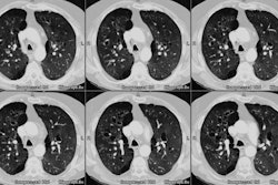
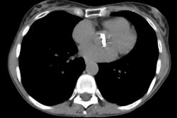
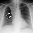
OBJECTIVE: In the setting of blunt trauma, abdominal CT, which routinely includes images of the lower thorax, frequently reveals pneumothoraces that have not been detected on routine supine chest radiographs. Proper management of these occult pneumothoraces remains controversial. The purpose of this study was to test the hypothesis that small (minuscule) to moderate (anterior) radiographically occult pneumothoraces can be safely managed without chest tube placement for patients in whom the need for positive pressure ventilation is not anticipated. SUBJECTS AND METHODS: We undertook a prospective study in which 44 occult pneumothoraces were classified into three groups, minuscule, anterior, or anterolateral, according to size and location on CT scans. Choice of initial management (tube thoracostomy versus close observation) was based in part on this classification system and in part on individual circumstances of a surgeon's decision. RESULTS: Of the 44 pneumothoraces found in 36 patients, 16 pneumothoraces were minuscule, 20 were anterior, and eight were anterolateral. Thirteen minuscule pneumothoraces and 11 anterior pneumothoraces initially managed with observation did not require subsequent tube thoracostomy. All eight patients with anterolateral pneumothoraces underwent tube thoracostomy. CONCLUSION: Most small (minuscule) occult pneumothoraces can successfully be managed with close observation. The risk that the pneumothorax will progress is slight. Moderate-sized (anterior) pneumothoraces may also be successfully managed without initial placement of a chest tube if the patient is not to undergo positive pressure ventilation.
