Post-pneumonectomy Complications:
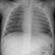
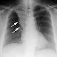
The overall incidence of complications after pneumonectomy has been reported to be as high as 20-60% [9]. The mortality rate following pneumonectomy is approximately 6%, which is three times higher than after lobectomy. Complications are more common following right pneumonectomy than left. The major causes of death are pneumonia (mortality 25%), pulmonary edema, pulmonary embolism, myuocardial infarction, empyema, and bronchopleural fistula.Initially, chest radiographs should demonstrate a midline position of the trachea, slight congestion of the remaining lung, and a postpneumonectomy space that contains gas and fluid [9]. In the normal post-pneumonectomy chest the potential pleural space is replaced by liquid from bleeding, weeping from lymphatics, and passive transudation. Within the first 4-5 post operative days, approximately half of the space becomes filled with fluid [9]. Gas is usually resorbed in 7 to 14 days, but the process can require up to 6 months and in some patients a small amount of gas persists indefinitely. Rapid opacification of the potential pleural space may reflect hemorrhage. Chylothorax secondary to thoracic duct injury can also occur. The mediastinum and heart gradually shift toward the side of the pneumonectomy and the remaining lung will expand across the mid-line. A contralateral mediastinal shift of up to 3.5 cm with expiration is acceptable. On long term follow-up, CT demonstrates that approximately two-thirds of patients maintain a liquid filled space with thick fibrous margins, while the remaining patients have fibrous tissue or normal mediastinal structures. [2] In the late postoperativ eperiod, a mediastinal shift toward the remaining lung is indicative of a delayed complication [9].
Lung torsion
Cardiac herniation/torsion
Hemothorax:
Major hemorrhage is most commonly due to inadequate hemostasis of a bronchial artery or systemic vessel in the chest wall [6]. It appears as a rapidly enlarging pleural fluid collection.
Chylothroax:
Chylothorax can occur secondary to thoracic duct injury. The attenuation on CT imaging can be variable- it may be low due to the fat content, but it can be higher because of a high protein content within the fluid [6].
Persistent air leak:
This complication is associated with incomplete or absent interlobar fissures and underlying emphysema [6]. It is defined as an air leak which persists more than 7 days [6].
Bronchopleural Fistula (BPF):
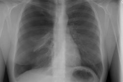
Bronchopleural fistula is a potetnial fatal complication of pneumonectomy [9]. The incidence of bronchial stump leak is 5% with a mortality rate of 16-23%. It is more common following right pneumonectomy- possibly related to features of the right main bronchus such as larger size, greater tendency to spring open, less mediastinal coverage of the stump, and a single bronchial artery which raises the vulnerability for ischemia [6,9]. Other factors associated with BPF include mechanical ventilation, preoperative radiation therapy, and preoperative pleuropulmonary infection [6]. Leakage during the first week is less common, likely due to poor closure, and requires re-operation. Leakage in the second or third week is usually due to poor healing. A short stump and preservation of the bronchial artery or peribronchial tissue can aid in decreasing the risk of stump necrosis and BPF formation [6]. The diagnosis is usually confirmed with bronchoscopy. Radiologic signs of BPF include: the appearance of an air-fluid level in a previously opaque post pneumonectomy space, failure of the potential pleural space to fill with liquid, inspiratory shifting of the mediastinum to the contralateral (non-operated) side, progressive mediastinal emphysema, and an abrupt decrease in the gas-liquid level greater than 2 cm in height. CT may demonstrate the tract from the airway into the pleural space [6,9]. [2,6] A decrease in fluid height of less than 1.5 cm may be due to differences in posture or in the degree of inspiration and should not be considered abnormal unless accompanied by mediastinal shift away from the postpneumonectomy space [9].Post-pneumonectomy empyema:
Post-pneumonectomy emyema occurs in 2-16% (incidence is probably less than 5% [9]) of patients and has a mortality rate between 16-71% [6]. BPF is the main cause of post-pneumonectomy empyema formation (about 5% of empyemas are associated with bronchopleural fistulas) [6]. On imaging studies, empyema appears as multiple air-fluid levels with a diffusely enhancing pleura [6]. There will be a shift toward the side opposite the pneumonectomy [9].
Pulmonary Edema:
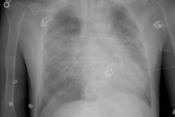
Post-pneumonectomy pulmonary edema is a life threatening complication that can result from volume overload or abrupt hyperperfusion- typically within 24 to 48 hours after surgery. It occurs in 1-5% of post-pneumonectomy patients (and in less than 1% of post-lobectomy patients) [4,6]. Predisposing factors include as excessive perioperative fluid load, transfusion of fresh frozen plasma, arrhythmia, and marked post surgical diuresis [9]. The condition occurs more commonly after right pneumonectomy (the remaining left lung has a smaller volume and must now handle the total pulmonary blood flow) and should be treated aggressively with diuretics. [2,9] The mortality rate can be as high as 80-100% [9].Post-pneumonectomy Syndrome:
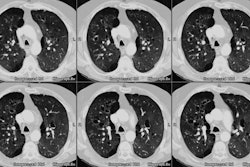
Post-pneumonectomy syndrome is a rare, delayed complication of right pneumonectomy (rarely left pneumonectomy in patients with right aortic arches). The syndrome occurs secondary to severe mediastinal shift which results in counterclockwise rotation of the heart and great vessels, and herniation of the remaining lung into the contralateral hemithorax. The trachea and remaining main bronchus can be compressed by the thoracic spine, descending aorta, ligamentum arteriosum, or pulmonary artery. Patients present with exertional dyspnea, inspiratory stridor, and recurrent infections in the remaining lung [9]. It seems to be more common in women and young children- possibly due to increased elasticity and compliance of their lungs and mediastinum [9]. CT images show abnormal narrowing of the distal part of the trachea and left main bronchus because of compression of the airway between the pulmonary artery anteriorly and the aorta and spine posteriorly [9]. Surgical repositioning of the mediastinal structures can be curative. [1,2]Stump Clot (in situ thrombus):
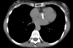
Stump clot is not uncommon (up to 12% of patients) and is likely related to stasis of blood flow [7,8]. The longer the stump, the greater the risk for clot formation [7,8]. Clot is more frequent on the right due to a longer stump [7]. The main risk is for propagation or migration into the remaining pulmonary circulation which could be fatal [3], however, this occurs only rarely [5,7,8].Pneumonia:
Pneumonia occurs in 2-15% of patients after pneumonectomy and is associated with a mortality rate of 25% [9].
REFERENCES:
(1) J Thorac Imag 1997; 12: 209-211
(2) AJR 1997; Tsukada G, Stark P. Post-pneumonectomy complications. 169: 1363-1370 (No abstract available)
(3) Radiology 1999; Remy-Jardin M, Remy J. Spiral CT angiography of the pulmonary circulation. 212: 615-636
(4) Radiographics 1999; Gluecker T, et al. Clinical and radiologic features of pulmonary edema. 19: 1507-1531
(5) AJR 2001; Wechsler RJ, et al. CT of in situ vascular stump thrombosis after pulmonary resection for cancer. 176: 1423-25 (No abstract available)
(6) Radiographics 2002; Kim EA, et al. Radiographic and CT findings in complications following pulmonary resection. 22: 67-86
(7) AJR 2005; Kim SY, et al. Filling defect in a pulmonary arterial stump on CT after pneumonectomy: radiologic and clinical significance. 185: 985-988
(8) Radiology 2005; Kwek BH, Wittram C. Postpneumonectomy pulmonary artery stump thrombosis: CT features and imaging follow-up. 237: 338-341
(9) Radiographics 2006; Chae EJ, et al. Radiographic and CT findings of thoracic complications after pneumonectomy. 26: 1449-1467
