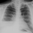
Effusion, Pleural:
Clinical:
The pleural space normally contains between 7-14 mL of fluid [14]. Pleural effusions may be transudative or exudative. Transudates result from an increase in the capillary hydrostatic pressure or a fall in the colloid osmotic pressure [14]. Common etiologies of transudative effusions include cardiac failure, constrictive pericarditis, cirrhosis, hypoproteinemia, and nephrotic syndrome.
An exudative effusion is the result of an increase in microvascular permeability- typically secondary to infection, infarction, or tumor [14]. An effusion is classified as an exudate if it meets any one of the following (Light's criteria): 1- the ratio of pleural fluid protein to serum protein is greater than 0.5; 2- the ratio of pleural fluid LDH to serum LDH is greater than 0.6; or 3- the pleural fluid LDH is greater than 200 IU. Exudates also have a specific gravity greater than 1.016 and a protein level greater than 3 g/dL.
Thoracentesis carries small, but significant risks with major
complication rates ranging from 12-30% [14]. The most common
complication is pneumothorax- occuring in around 12% of procedures- and
5% of patients require chest tube insertion [14]. Relative
contraindications to thoracentesis include bleeding diathesis,
anticoagulation, mechanical ventilation, and overlying cutaneous
disease or infection [14].
Re-expansion pulmonary edema can result from the rapid removal of
air or fluid from the pleural space, usually after prolonged pulmonary
atelectasis [17]. It is thought to be related to increased pulmonary
vascular permeability, surfactant depletion, and increased production
of oxygen free-radicals [17]. The clinical maifestations can vary from
minimal symptoms to severe hypoxemia and cardiorespiratory collpase
[17]. The symptoms tend to occur within the first 2 hours after lung
reexpansion, but may take up to 48 hours to appear [17]. The process
usually lasts 1-2 days, but can take longer to resolve [17]. The main
finding on CXR is a unilateral airspace opacity, which can be seen
within a few hours of re-expansion of the lung [17]. CT findings
include ground-glass opacities, consolidation, and inter/intralobular
septal thickening [17].
Parapneumonic Effusion:
A pleural effusion associated with a pulmonary infection is known as a parapneumonic effusion. Parapneumonic effusions occur in about 40% to 50% of patients with community acquired pneumonia [5,6]. Parapneumonic effusions may be simple or complicated. Simple parapneumonic effusions probably result from increased permeability of the visceral pleura. Although they are commonly exudates, simple parapneumonic effusions do not demonstrate frank pus, have normal glucose and pH values, and are culture and gram stain negative. Most simple parapneumonic effusions will resolve with appropriate antibiotic treatment of the underlying pneumonia.
Complicated parapneumonic effusions are gram stain or culture negative, but fluid analysis will demonstrate a high LDH level (over 1000 IU/L), a low pH (less than 7.2), or a low glucose level (less than 40 mg/dL). A complicated parapneumonic effusion requires chest tube drainage [1]. Radiologic guided chest tube drainage is most effective in patients with short duration of symptoms, free-flowing or unilocular fluid collections, and no evidence of a thick pleural peel on CT scan [5]. If not drained, complicated parapneumonic effusions will become loculated with a significant fibrotic response within the pleural space [6]. Percutaneous drainage is of limited value at this stage [6]. The fibrosis can produce significant morbidity with impaired respiratory motion [6].
Click here for a discussion of Empyema
Malignant Effusion:
About 60% of patients with metastatic disease to the pleura will develop pleural effusion- typically as a result of mediastinal lymphatic obstruction. Malignant effusion is most commonly secondary to bronchogenic carcinoma, following by breast cancer, and lymphoma. Between 90-95% of malignant effusions are exudative. Pleural metastases commonly involve both the parietal and visceral pleura. Pleural involvement almost always indicates a poor prognosis. Generally, malignant effusions are relatively large (500-200 ml). Cytologic evaluation of the fluid will be positive for malignancy in about half to two-thirds of cases. Repeated thoracentesis has been reported to identify an additional 30% of patients with malignant effusion and is recommended prior to pleural biopsy [9]. Reverse-bevel needle biopsy of the pleura is accurate in only 48-60% of patients with malignant effusions, and is also not sensitive for the detection of mesothelioma (sensitivity 21-43%) [9]. CT guided biopsy can increase the sensitivity for the diagnosis of malignancy to 72-97% [10]. Accuracy of greater than 95% can be achieved by using thoracoscopic guided biospy [9].
Patients with malignant effusions frequently require therapeutic thoracentesis for symptomatic relief of dyspnea. A subgroup of these patients will develop asymptomatic hydropneumothorax following thoracentesis due to decreased lung compliance with failure of re-expansion (pneumothorax ex vacuo). Causes of poor lung compliance include fixed atelectasis (from endobronchial obstruction), lymphangitic metastases, and visceral pleural metastases. Chest tube drainage of the pneumothorax is not required as the pleural fluid will reaccumulate over a variable period of time [7].
Clinical management of patients with recurrent malignant effusions may involve pleurodesis (pleurodesis is the palliative treatment of choice for recurrent malignant pleural effusion). For pleurodesis a sclerosing agent is instilled into the pleural cavity (chemical pleurodesis) or there is surgical abrasion of the pleura with a dry gauze sponge (mechanical pleurodesis) [13]. Complete pleural fluid evacuation with reexpansion of the underlying lung and apposition of the visceral and parietal pleural surfaces is necessary for successful pleurodesis. A thick pleural peel, endobronchial obstruction, or underlying interstitial lung disease can all prevent reexpansion and preclude a successful outcome [5].
Sclerosing agents include talc, doxycycline, minocycline, and bleomycin. The patient is rotated to ensure adequate distribution of the agent. Talc is the most common sclerosing agent used for pleurodesis. It can be administered as a slurry via chest tube or by poudrage during thoracoscopy [13]. Talc produces a visceral and parietal pleural granulomatous inflammation. Thoracoscopic installation of talc power has been successful in 85-90% of patients, but the procedure requires that the patient undergo general anesthesia. Complications from talc pleurodesis include adult respiratory distress syndrome and an increased risk for mesothelioma [13]. The risk for ARDS is dependent on the dose of talc administered and is almost completely eliminated by using a dose of 2 g or less of talc [13]. The risk for mesothelioma was previously attributed to asbestos in unpurified talc [13]. The talc now used for pleurodesis is purified and free of asbestos [13] which should eliminate the risk for secondary malignancy. Although on follow-up chest radiographs the talc deposits are not typically seen, they can be identified as clusters of high attenuation on CT images [2]. The pleural talc deposits are most commonly seen in the posterior caudal regions of the pleural space [11]. Increased FDG activity has been reported in sites of pleural talc (up to 3 years following the procedure [11])- presumably secondary to pleural inflammation [3,11].
Bleomycin or other chemical agents are mixed with lidocaine and administered via a pleural tube. Bleomycin instillation into the pleural space can prevent recurrent effusion in 60-90% of patients for up to one month. Adverse side effects include nausea, vomiting, bone marrow suppression, rash, and diarrhea [13]. The high cost of the agent has also limited its use [13]. Doxycycline is now also commonly used, but it may require multiple installations (in up to 72% of patients) to be effective [13].
Pleuroperitoneal shunting has also been used in some patients. [4]
Other etiologies of pleural effusions:
Hepatic Hydrothorax:
Hepatic hydrothorax is defined as a significant pleural effusion usually greater than 500 mL in a cirrhotic patient in the absence of cardiopulmonary disease [15]. Up to 10% of patients with significant liver disease develop pleural effusions [8]. The pressure gradient favors the movement of fluid from the peritoneal cavity to the pleural space through small diaphragmatic defects. The effusion is right sided in 67-85% of cases, bilateral in 2-17%, and left sided in 13-17% [8,15]. Hepatic hydrothorax may occasionally occur in the absence of ascites [15].
X-ray:
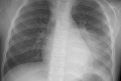
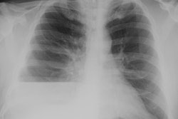
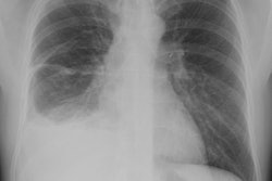
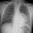
CXR: On a PA CXR, approximately 200 ml of pleural fluid will produce lateral CP angle blunting and a volume of 500 mL will usually obscure the diaphragm [12]. On the lateral exam, about 50 mL is the smallest amount of fluid to produce posterior CP angle blunting [12]. However, a lateral decubitus film can demonstrate as little as 5 ml of fluid [12].
Computed tomography: HU numbers cannot be used to reliably distinguish a transudative from an exudative effusion. The presence of a thickened partietal pleura on contrast exam indicates that the effusion is an exudate, however, the absence of this enhancement does not exclude an exudate. Pleural enhancement is identified in 61% of patients with exudative effusions and is more common in infection than in malignancy. Loculated effusion is also more common with exudates [14].
Approximately 50% of malignant effusions resemble simple transudative effusions without pleural changes [12]. Pleural thickening or nodularity is seen in association with about 30% of malignant effusions. PET FDG has been shown to be abnormal in patients with malignant pleural effusions [9]. Increased pleural FDG uptake is highly suggestive of a malignant effusion (sensitivity 95%) [9].
Distinction of pleural from peritoneal fluid may sometimes be difficult. CT findings which permit differentiation of pleural fluid from ascites include: The diaphragm sign- pleural fluid is outside the diaphragm, ascites inside; the interface sign: ascites is usually of lower attenuation than the liver, spleen, or hemidiaphragm and the interface is sharp, whereas it is ill-defined with a pleural effusion; the bare area sign: the bare area is the portion of the right lobe of the liver which lacks a peritoneal covering. In this area the liver is directly attached to the posterior abdominal wall and ascites cannot extend behind the liver at this level; and the displaced crus sign- which refers to anterior displacement of the diaphragmatic crus by pleural fluid.
REFERENCES:
(1) Radiographics 1997; 17: 63-79
(3) AJR 1997; Murray JG, et al. Talc pleurodesis simulating pleural metastases on 18F-fluorodeoxyglucose positron emission tomography. 168: 359-360 (No abstract available)
(4) ACR Syllabi #40: p.40-42; 142-45
(6) J Thorac Imaging 1998; Patz EF, et al. Percutaneous drainage of pleural collections. 13: 83-92
(7) AJR 1998; Boland GW, et al. Asymptomatic hydropneumothorax after therapeutic thoracentesis for malignant pleural effusions. 170: 943-946
(8) Radiographics 2000; Meyer CA, et al. Diseases of the hepatopulmonary axis. 20: 687-698
(9) AJR 2000; Erasmus JJ, et al. FDG PET of pleural effusions in patients with non-small cell lung cancer. 175: 245-249
(10) Radiology 2001; Adams RF, Gleeson FV. Percutaneous image-guided cutting-needle biopsy of the pleura in the presence of a suspected malignant effusion. 219: 510-514
(11) AJR 2004; Nguyen M, et al. CT with histopathologic correlation of FDG uptake in a patient with pulmonary granuloma and pleural plaque caused by remote talc pleurodesis. 182: 92-94
(12) Radiol Clin N Am 2005; Aquino SL. Imaging metastatic disease to the thorax. 43: 481-495
(13) AJR 2006; Avila NA, et al. CT of pleural abnormalities in lymphangioleiomyomatosis and comparison of pleural findings after different types of pleurodesis. 186: 1007-1012
(14) AJR 2009; Abramowitz Y, et al. Pleural effusion: characterization with CT attenuation values and CT appearance. 192: 618-623
(15) Radiographics 2009; Kim YK, et al. Thoracic complications of
liver cirrhosis: radiologic findings. 29: 825-837
(16) AJR 2012; Godoy MCB, et al. Chest radiography in the ICU part
I, evaluation of airway, enteric, and pleural tubes. 198: 563-571
(17) AJR 2012; Godoy MCB, et al. Chest radiography in the ICU part I, evaluation of airway, enteric, and pleural tubes. 198: 563-571
