Bronchiolitis obliterans syndrome: thin-section CT diagnosis of obstructive changes in infants and young children after lung transplantation.
Lau DM, Siegel MJ, Hildebolt CF, Cohen AH
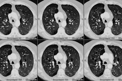
PURPOSE: To characterize the thin-section computed tomographic (CT) appearance of bronchiolitis fibrosa obliterans syndrome in infants and young children after lung transplantation. MATERIALS AND METHODS: Thin-section CT studies in six patients with bronchiolitis obliterans syndrome (age range, 2 months to 5 1/2 years) and in 15 control patients without obstructive airway disease (age range, 2 months to 7 years) who underwent bilateral lung transplantation were retrospectively reviewed. The thin-section CT scans were obtained during quiet sleep at a median of 24 months (range, 6-36 months) after transplantation. The CT studies were evaluated for mosaic perfusion, bronchial dilatation, bronchial wall thickening, and mucous plugging Final diagnoses in all patients were based pulmonary function test results. RESULTS: Thin-section CT findings in the six patients with clinically proved bronchiolitis obliterans syndrome were mosaic perfusion in five (83%) bronchial dilation in three (50%), and bronchial wall thickening in one (17%). Of the 15 control patients with normal pulmonary function test results, six (40%) had mosaic perfusion; none had bronchial dilatation or bronchial wall thickening. Mucous plugging was not seen in either group. Only the association of bronchial dilatation with bronchiolitis obliterans syndrome was significant (P = .02). CONCLUSION: Infants and young children with bronchiolitis obliterans syndrome after lung transplantation are more likely to have CT abnormalities than those with normal pulmonary function test results.
