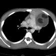
Thymic Lesions:
The thymus arises from the third and fourth branchial pouches and contains elements from all three germ cell layers [15]. The gland consists of two lateral lobes placed in close contact along the midline of the upper chest [14]. The thymus increases from birth to between 4 and 8 months of age and then decreases [14]. It normally weighs about 15 gm at birth and 35 gm at puberty [14]. It is most active and largest during puberty after which it shrinks in size and activity in most people and is replaced with fat [14]. Total fatty involution of the thymus occurs around the age of 40 years when only about 5% of residual thymic tissue is retained [19]. However, the normal thymus can still be identified in up to 100% of patients under the age of 30 years, in up to 73% of patients between the ages of 30-49 years, and in up to 17% of patients older than 49 years [13].
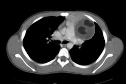
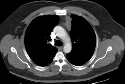
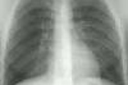
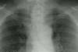
On CXR in infants and young children the thymus is strikingly large- it usually has smooth borders and remains visible on radiographs through 3 years of age [20]. A scalloped or wavy contour (the thymic wave sign) occurs secondary to anterior rib impressions on the thymus [20]. The thymic sail sign - a triangular slightly convex right lobe of the thymus with a sharply demarcated base produced by the minor fissure is seen in about 5% of children [20]. On non-contrast CT the thymus appears similar in density to muscle tissue [14]. After contrast administration the thymus shows homogeneous enhancement of 20-30 HU [14]. Thymic morphology varies greatly- in young adults it is typically bilobed and V-shaped with two small processes extending into the neck [20]. On MR, the normal thymus has homogeneous signal on T1 which is slightly brighter than muscle [14]. On T2, the signal becomes much brighter than muscle [14]. On opposed phase chemical shift imaging the normal thymus and hyperplastic thymus have been shown to demonstrate decreased signal intensity due to the presence of fat within the tissue [13]. Thymic heterogeneity on fat saturation sequences should be considered pathologic [14]. Gallium 67 will accumulate in the normal thymus in up to 60% of children under 2 years of age and also in activated thymic tissue following chemotherapy and systemic illness [14]. I-131 uptake has also been reported in hyperplastic thymic tissue that does not contain ectopic thyroid tissue [14]. The thymus can also be visualized on Octreotide imaging due to the presence of somatostatin receptors in the normal thymus [14]. The normal thymus can accumulate FDG and be visualized on PET imaging [14].
- Thymoma
Thymoma / Invasive Thymoma:
View cases of thymoma
Clinical:
Thymomas generally occur in patients over the age of 30 years abd are rare in children (70% of lesions are found in adults between the 5th and 6th decades) [12,21]. Thymomas occur with equal frequency in men and women [20,21]. Approximately 35% of thymomas are invasive, but they are usually indistinguishable from benign tumors radiographically. Most patients are asymptomatic (20-50% of patients [12]), however, thymomas are associated with myasthenia gravis. About 35-40% of patients with thymomas have myasthenia, whereas 10-15% of myasthenia patients have a thymoma [21]. Thymomas associated with myasthenia are typically less aggressive and have a better prognosis. Thymomas are also associated with other hematologic disorders such as hypogammaglobulinemia (Good's Syndrome-in 5% of cases) and red cell aplasia (in about 5%- particularly spindle cell thymoma). Conversely, about 10% of patients with hypogammaglobulinemia are discovered to have a thymoma, while about 50% of patients with red cell aplasia have a thymoma. Thymectomy for thymoma frequently alleviates the symptoms of myesthenia gravis, but only occasionally results in remission of red cell aplasia and it never improves hypogammaglobulinemia [6,8].
Thymomas rarely metastasize and more commonly they extend locally
or seed the pleural or pericardial surfaces. Thymic epithelial
tumors consist of several histologic subtypes in order of
increasing malignancy- A, AB, B1, B2, B3, and thymic carcinoma
[16]. Thymoma types A and AB generally behave like benign tumors,
B1 is a low-grade malignant tumor (over 90% ten year survival), B2
has a higher degree of malignancy, and B3 has a poor prognosis
[16]. In general, A, AB, and B1 are considered low risk lesions,
while B2 and B3 are high risk [16].
Histologic differentiation of benign from invasive lesions is not possible and lesions are termed "invasive" if there is evidence of extension of the tumor beyond its surrounding fibrous capsule at histologic analysis. Although patients with encapsulated thymomas have the best prognosis and are almost always cured with complete excision, 2-12% of resected encapsulated thymomas recur, sometimes months or years later [5]. The five year survival for patients with encapsulated thymoma is about 75%, while those with invasive thymomas have a 50% 5 year survival [5]. After surgery, radiation therapy is recommended to reduce the risk of recurrences in patients with invasive thymomas (typically 40-60 Gy) [12]. About 5% of patients with invasive thymomas have distant metastases [20]. Patients with invasive thymomas who develop metastases and all patients with stage IV disease are candidates for chemotherapy (cisplatin- overall response rate 40-90%) [12]. Because of the risk of recurrence, all thymomas warrant long-term follow-up. The presence of coexistent myasthenia does not adversely affect patient survival [8].
Current Masaoka-Koga staging for thymomas (from Masaoka et al,21):
Stage I- tumor with intact capsule; Stage II- transcapsular
extension into the mediastinal fat (IIa- microscopic invasion
through the capsule and IIb- macroscopic invasion into surrounding
fat); Stage III- invasion of the adjacent organs - focal pleural
or pericardial, mediastinal structures (great vessels, heart), or
lung; Stage IVa- pleural or pericardial dissemination; and Stage
IVb- Lymphatic or hematogenous metastases [21]. Stage I lesions
are treated with surgical resection alone, while stage II lesions
are treated with surgery and post op XRT if the resection is
incomplete [21]. For stage III and IVa lesions, patients receive
adjuvant chemotherapy followed by surgery and XRT if required
[21]. Stage IVb lesions are treated with palliative chemotherapy
[21].
Prognosis for a non-invasive thymoma is very good with only a 2% recurrence rate for encapsulated lesions. Invasive lesions have a 50-78% 5 year survival [2]. The reported average time to recurrence of a completely resected thymoma is approximately 5 years- hence, long term followup of these patients is required [21].Excision of the lesion generally results in improvement in the clinical status of myasthenia patients- often resulting in complete remission.
On histologic analysis a thymoma consists of a mixture of thymic epithelial cells and lymphocytes. The epithelial cells may be spindle-shaped and if more than two thirds of the epithelial cells have this appearance, the thymoma is classified pathologically as a 'spindle cell tumor.' Purely lymphocytic and spindle cell thymomas have been suggested to be less aggressive than epithelial and mixed lymphocytic/epithelial thymomas [3].
X-ray:
Between 25% to 50% of thymomas are undetectable on PA chest radiographs [6,12]. CT scan is the study of choice in the evaluation of patients with myesthenia gravis or a suspected thymoma [12]. CT has a sensitivity of 91% and a specificity of 97% for the diagnosis of a thymic mass [12]. MR adds little additional information. Note: The use of iodinated contrast agents in patients with myasthenia gravis has been reported to cause increased weakness. Although not contraindicated, intravenous contrast agents should not be used routinely in the assessment of these patients. [1]
On CT, thymomas appear as a lobulated, well-defined, soft tissue density mass with rounded margins in the anterior mediastinum and growth is usually to one side. Mild, homogeneous contrast enhancement is characteristic. Calcification is common (20% of cases on plain film). Cystic areas (due to hemorrhage or necrotic degeneration) are also found in 20-30% and these are typically larger lesions. A clue to the diagnosis is that a thymoma will be surrounded by fat, while the normal thymus is diffusely infiltrated with fat. Partial obliteration of the mediastinal fat planes about the lesion does not confirm the diagnosis invasion (finding may be seen in about 50% of patients with non-invasive lesions) [2]. However, an indistinct border between a thymoma and the adjacent lung or the presence of a pleural effusion or pleural nodularity suggest invasion [20]. Spread to periaortic nodes in the upper abdomen has been reported in up to 31.5% of patients with invasive thymomas.
On MR, thymomas commonly appear as homogeneous or heterogenenous
masses with low signal on T1 and high signal on T2
[20].Differentiation from non-neoplastic thymic enlargement can be
performed with chemical shift MR imaging [21]. This technique
allows identification of the normal fatty infiltration of the
thymus that will manifest with homogeneous decreased signal on the
out-of-phase images [21].
Thallium imaging has also been used to evaluate patients [12]. The normal thymus demonstrates no increased thallium concentration, whereas lymphoid follicular hyperplasia shows moderate thallium uptake on delayed images, and thymoma shows moderate to strong thallium uptake on both early and delayed images [12].
Thymic Carcinoma:
Clinical:
Thymic carcinoma is an uncommon tumor and it is only very rarely association with autoimmune/paraneoplastic disorders [9]. Unlike an invasive thymoma which has histologic features that are indistinguishable from a benign thymoma, this lesion shows true malignant features [9]. Various histologic subtypes have been described including- squamous cell (36%), lymphoepithelioma-like (32%), undifferentiated or anaplastic (11%), small cell, sarcomatoid, clear cell, and adenocarinoma [9]. The majority of patients are middle aged (avg. age 46-50 years) and there is a slight male predominance. Nearly one third of patients will be asymptomatic, while those with symptoms will complain of chest pain, cough, dyspnea, hoarseness, dysphagia, or SVC syndrome (13-25%). Non-specific systemic complaints are also found such as fever, night sweats, or weight loss. Unlike thymomas, thymic carcinomas rarely cause paraneoplastic syndromes such as myasthenia gravis [20]. The tumor is most commonly of the squamous cell variety. The lesion is not encapsulated and there is gross local infiltration. Lymphatic and distant mets are common (55-65% of patients at time of diagnosis [20]). The presence of distant metastases helps to differentiate the lesion from an invasive thymoma which tends to spread locally, and not hematogenously [9]. The lesion may respond to treatment with chemotherapy, but patients have an overall very poor prognosis with an average 5 year survival of 10-65%.
X-ray:
On CT the tumor is usually inhomogeneous due to internal necrosis and hemorrhage. Many of the tumors demonstrate gross local invasion of contiguous mediastinal structures (80% of cases [8]) and mediastinal lymphadenopathy is seen in 40% [8]. Calcifications can be seen within the lesion. Unfortunately, there are no distinctive CT features which permit differentiation of thymic carcinoma from an invasive thymoma unless the presence of hematogenous metastases can be demonstrated. [2]
Thymic Carcinoid/Neuroendocrine Neoplasm:
Clinical:
Thymic carcinoid is very rare (0.4% of all carcinoids arise from
the thymus [27]), low-grade malignant lesion with an aggressive
behavior typically seen in middle aged patients (30-60 years;
median age at presentation 57 years) [17,20,26,27]. Men are
affected more than women (3:1) [7,8,15,17]. The majority of
patients present with symptoms related to mass effect or invasion
of mediastinal structures, but about one-third are asymptomatic
[7]. About 50% of thymic carcinoids are invasive at the time of
diagnosis and approximately 20% of patients have metastases at
presentation [8,20]. The most frequent sites for metastatic
disease are regional lymph nodes (lymph node involvement can be
seen in up to 50% of patients at diagnosis [27]), the adrenal
glands, and bone [17]. Pulmonary, pleural, cerebral, and renal
metastases occur less commonly [17]. About 33-50% of thymic
carcinoids are functionally active [8,15], but do not produce
carcinoid syndrome [8].
Cushing's syndrome (hypercortisolism due to ectopic ACTH
production) occurs in 33-40% of cases [7]. Interestingly, the 10
year mortality rate is higher (65%) when associated with Cushing
syndrome, compared to 29% in patients with no associated
endocrinopathy [26].
The lesion is associated with Wermer's syndrome (MEN I- hyperparathyroidism, islet cell tumors of the pancreas, and pituitary adenomas) which is seen in 19-25% of patients with thymic carcinoids (conversely, thymic neuroendocrine neoplasms develop in 3-8% of MEN I patients [27]) [7,17]. The lesion tends to be more aggressive in these patients [8] and there may be an association with heavy smoking [27]. It can also be seen in patients with MEN II [20]. Complete excision can be curative, but local recurrence or mets develop in 70% of cases. Adjuvant radiation and chemotherapy are often employed. Prognosis is poor with a 5-year survival rate of about 30% [7].
On CT the lesion appears as a very vascular enhancing, solid anterior mediastinal mass. The lesion can be localized or invasive. Calcifications and small areas of necrosis may be seen. [3] Scintigraphy with Octreotide will accumulate within the lesion, but the finding is non-specific as the tracer will also accumulate in other thymic tumors and lymphoma.
Thymolipoma:
Clinical:
Thymolipoma is a rare, slow growing benign well-encapsulated tumor composed of thymic tissue and fat (probably best classified as a hamartoma). Affected patients present with a mean age of approximately 30 years and men and women are affected equally. The lesion is non-invasive and only rarely associated with myasthenia gravis. Most patients are asymptomatic [20]. It is a very soft, deformable tumor that will conform to adjacent structures and is often quite large by the time of presentation (mean diameter 20 cm) [20]. Histologically the tumor consists of both normal thymic and adipose tissue. The tumor has a thin capsule and surgical excision is curative. There are no reports of malignant degeneration [8].
X-ray:
On CXR the lesion appears as a mass or mediastinal widening that has sharp margins with the adjacent lung. The lesion frequently mimics an enlarged cardiac silhouette on the frontal radiograph. Because the lesion is soft, it tends to drape over the ipsilateral hemidiaphragm, conforming to its shape and simulating diaphragmatic elevation on the lateral radiograph. The lesion may change shape with change in patient position. The failure of the lesion to silhouette the diaphragm helps to confirm its fat content. On CT the lesion arises from the anterior mediastinum and contains areas of fat density (90% of cases) with variable amounts of soft tissue thymic stroma. This thymic tissue often has the appearance of linear whorls [10]. Fat usually constitutes for between 50-90% of the lesion [10]. Differential considerations include lipoma/ liposarcoma, mature teratoma, or omental herniation. [4]
Thymic Cyst:
Clinical:
Thymic cysts are uncommon lesions that can be congenital or acquired [8]. Congenital thymic cysts are rare and derive from a patent thymopharyngeal duct [11]. The thymus arises from the 3d and 4th pharyngeal pouches. Remnants of these pouches in the neck can form well encapsulated cysts that are lined by epithelium. These lesions generally arises from one side of the gland and present in childhood (about 50% of lesions are discovered incidentally during the first 2 decades of life [11]). Most are found in the anatomical location of the thymus, but they can be found anywhere along the course of thymic descent from the neck into the mediastinum [8].
Most thymic cysts are not discovered until adulthood- most likely because the lesion is typically asymptomatic and discovered on routine CXR. Although possibly congenital, thymic cysts in adults are likely acquired lesions and represent the sequella of prior inflammation or radiation therapy [8,11]. Thymic cysts have also been described in HIV patients [8]. Surgical excision is curative.
Thymic cysts usually appear as a well circumscribed mass in the anterosuperior mediastinum. CT attenuation measurements are usually near water density and the cyst wall is typically thin or invisible. Internal septa or mural calcification may occur [8].
Acquired thymic cysts have been reported to occur before and
after chemotherapy for non-Hodgkin or Hodgkin lymphoma, after
thoracotomy, and in about 40% of patients with thymomas [20].
HIV stimulates the proliferation of CD8 T lymphocytes that results in a set of diseases known as diffuse infiltrative lymphocytosis syndrome [23]. These diseases include LIP in the lungs, lymphoepithelial cysts in the salivary glands, and multilocular thymic cysts [23]. Multilocular thymic cysts in children with HIV infection were reported prior to retroviral therapy that is now available [20].
X-ray:
On CT the lesion appears as a near water density mass with imperceptible walls, but the lesion may appear of increased density if complicated by hemorrhage or infection. The lesion can be uni- or multilocular [11]. Dystrophic curvilinear calcification may be noted within the wall of complicated cysts. The lesions is usually located near the base of the heart.
Thymic Hyperplasia:
Clinical:
There are two forms of thymic hyperplasia:
1- True thymic hyperplasia (or rebound hyperplasia), in which
there are normal proportions of glandular/ lymphoid elements in an
enlarged gland secondary to rebound following stress-related
atrophy, illness, or immune suppression (following chemotherapy or
discontinuation of corticosteroid administration) [15,19]. When
the body is exposed to stress the thymus may shrink to as little
as 40% of its original volume (depending on the severity and
duration of the stress) [20]. Once the body recovers, the thymus
can usually grows back to its original size within 9 months, but
it can grow to be as much as 50% larger [20]. Approximately 10-25%
of patient who undergo chemotherapy may develop rebound
hyperplasia [20]. Most cases of thymic hyperplasia secondary
to chemotherapy occur within a year or two and the gland typically
returns to normal size [8,20]. Because of bolstered cell-mediated
immunity, thymic hyperplasia has been associated with a good
prognosis in post-chemotherapy patients [19]. Thymic hyperplasia
has also been described in patients with endocrine abnormalities
such as Graves' disease, acromegaly, and thyrotoxicosis [8], as
well as during recovery from burn injuries, in patients with
infections, and after cardiovascular surgery [19]. On CT the gland
may be normal sized or there may be symmetric enlargement of the
gland with preservation of its normal shape and no discrete mass.
The borders of the enlarged thymus are most often concave, but
occasionally the gland may have a convex margin and appear oval.
Streaks of fat should be identified and the gland should maintain
a bilobed appearance.
2- Thymic lymphoid follicular hyperplasia:
Thymic lymphoid follicular hyperplasia is characterized by the presence of an increased number of hyperplastic lymphoid follicles and germinal centers in the thymic medulla that is associated with lymphocytic and plasma cell infiltration [20,22]. It is more common than true hyperplasia and is associated with myasthenia gravis [20]. In fact, lymphoid hyperplasia is seen in 65-85% of patients with myasthenia. About 10-15% of myasthenia patients are discovered to have a thymoma, and about 20% have a normal or involuted thymus. Following thymectomy, a clinical remission occurs in 35% of myasthenia patients with lymphoid follicular hyperplasia, and 30-50% will demonstrate clinical improvement [8]. However, even myasthenia patients with a normal thymus may improve clinically following thymectomy (up to two-thirds of such patients). Thymic lymphoid hyperplasia is also associated with collagen vascular disorders such as scleroderma, rheumatoid arthritis, and SLE [15]; as well as endocrinopathies such as Graves disease [15], Addisons disease, acromegaly, and the early stages of human immunodeficiency virus infection [20].
On CT scan, thymic lymphoid hyperplasia can appear as an anterior mediastinal mass (20%), as a diffusely enlarged thymic gland (35%), or as a normal thymus (45%) [20]. It has been suggested that the non-contrast attenuation of the thymus is significantly higher in patients with lymphoid hyperplasia than in true hyperplasia (HU greater than 41.2) [22]. Note: The use of iodinated contrast agents in patients with myasthenia gravis has been reported to cause increased weakness. Although not contraindicated, intravenous contrast agents should not be used routinely in the assessment of these patients. [1]
Thallium may be beneficial in differentiating thymic hyperplasia from other thymic lesions. Normally, no thallium uptake should be seen in the mediastinum which appears photon deficient against the normal faint pulmonary activity. Therefore- the normal thymus is usually gallium avid, but will be negative on the thallium scan.
MR chemical shift imaging can also be used to differentiate
thymic hyperplasia from other thymic lesions [18,24]. Signal loss
on out-of-phase images compare to in-phase images suggests the
presence of fat and thymic hyperplasia, whereas neoplasms and
lymphoma do not demonstrate suppression on out-of-phase images
[18,22,24,25]. However, it should be noted that the amount of fat
tissue within the thymus increases with age [24].
FDG PET imaging is not useful for evaluation of thymic hyperplasia as the condition can be associated with FDG accumulation and there is considerable overlap with other thymic lesions [18].
REFERENCES:
(1) Radiology 1996; 201: 471-474
(2) J Thorac Imag 1996; 11: 66-74
(3) J Thorac Imag 1996; Abnormalities of the thymus.11:58-65 (No abstract available)
(4) Radiology 1994; Oct 193(1):121-126
(5) RadioGraphics 1998; Ros PR, et al. Image interpretation session: 1997. 18: 195-228 (Case1- pages 196-199) (No abstract available)
(6) AJR 1992; Fox MA, et al. Thymoma with hypogammaglobulinemia (Good's syndrome): An unusual cause of bronchiectasis. 158: 1229-1230 (No abstract available)
(7) Radiographics 1999; Rosado de Christenson ML, et al. Thoracic carcinoids: Radiologic-Pathologic correlation. From the archives of the AFIP. 19: 707-736
(8) J Thoracic Imag 1999; Strollo DC, et al. Tumors of the thymus. 14: 152-171
(9) AJR 2001; Jung KJ, et al. Malignant thymic epithelial tumors: CT-Pathologic correlation. 176: 433-439
(10) Radiographics 2002; Gaerte SC, et al. Fat-containing lesions of the chest. 22: S61-S78
(11) Radiographics 2002; Jeung MY, et al. Imaging of cystic masses of the mediastinum. 22: S79-S93
(12) Radiographics 2002; Santana L, et al. Best cases from the AFIP: Thymoma. 22: S95-S102
(13) AJR 2003; Takahashi K, et al. Characterization of the normal and hyperplastic thymus on chemical-shift MR imaging. 180: 1265-1269
(14) Radiol Clin N Am 2005; Franco A, et al. Imaging evaluation of pediatric mediastinal masses. 43: 325-353
(15) Radiographics 2006; Nishino M, et al. The thymus: a comprehensive review. 26: 335-348
(16) J Nucl Med 2006; Sung YM, et al. 18F-FDG PET/CT of thymic epithelial tumors: usefulness for distinguishing and staging tumor subgroups. 47: 1628-1634
(17) Radiographics 2007; Scarsbrook AF, et al. Anatomic and functional imaging of metastatic carcinoid tumors. 27: 455-476
(18) Radiology 2007; Inaoka T, et al. Thymic hyperplasia and thymus gland tumors: differentiation with chemical shift MR imaging. 243: 869-876
(19) J Nucl Med 2009; Jerushalmi J, et al. Physiologic thymic uptake of 18F-FDG in chidlren and young adults: a PET/CT evaluation of incidence, patterns, and relationship to treatment. 50: 849-853
(20) Radiographics 2010; Nasseri F, Eftekhari F. Clinical and
radiologic review of the normal and abnormal thymus: pearls and
pitfalls. 30: 413-428
(21) Radiographics 2011; Benveniste MFK, et al. Role of imaging n
the diagnosis, staging, and treatment of thymoma. 31: 1847-1861
(22) AJR 2014; Araki T, et al. Imaging characteristics of
pathologically proven thymic hyperplasia: identifying features
that can differentiate true from lymphoid hyperplasia. 202:
471-478
(23) Radiographics 2014; Chou SHS, et al. Thoracic diseases
associated with HIV infection in the era of anti-retroviral
therapy: clinical and imaging findings. 34: 895-911
(24) Radiology 2015; Priola AM, et al. Differentiation of rebound
and lymphoid thymic hyperplasia from anterior mediastinal tumors
with dual-echo chemical-shift MR imaging in adulthood: reliability
of the chemical-shift ratio and signal intensity index. 274:
238-249
(25) Radiographics 2017; Carter BW, et al. ITMIG classification
of mediastinal compartments and mutlidisciplinary approach to
mediastinal masses. 37: 413-436
(26) Radiographics 2017; Baxi AJ, et al. Multimodality imaging
findings in carcinoid tumors: a head-to-toe spectrum. 37: 516-536
(27) Radiographics 2017; Carter BW, et al. IASLC/ITMIG staging
system and lymph node map for thymic epithelial neoplasms. 37:
758-776
