AJR Am J Roentgenol 2000 Mar;174(3):789-93
Talcosis associated with IV abuse of oral medications: CT findings.
Ward S, Heyneman LE, Reittner P, Kazerooni EA, Godwin JD, Muller NL.
Department of Radiology, Vancouver General Hospital and University of British
Columbia, Canada.
OBJECTIVE: Our objective was to evaluate the CT appearance of talcosis
associated with IV abuse of oral medications and to compare the findings of
talcosis related to methylphenidate with those findings seen with other drugs.
MATERIALS AND METHODS: The CT scans of 12 patients with talcosis (seven men,
five women), 33-54 years old (mean age, 44 years), were analyzed
retrospectively. Seven patients had abused methylphenidate; five patients had no
history of abuse. The diagnosis of talcosis was made histologically in 11
patients and at funduscopy in one patient. CT was performed with 1- to 1.5-mm
collimation (n = 10 patients) or 5- to 10-mm collimation (n = 2). RESULTS: The
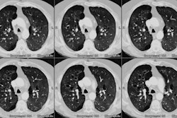
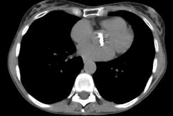
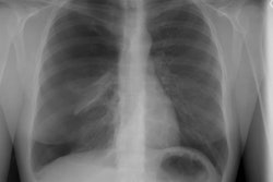
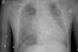
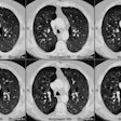
predominant abnormalities seen on CT consisted of a diffuse fine nodular pattern
(n = 2), a combination of nodules and lower lobe panacinar emphysema (n = 3),
and ground-glass attenuation (n = 2). Emphysema was the only abnormality seen in
the remaining five patients (lower lobe panacinar, n = 4; upper lobe
centrilobular, n = 1). No significant difference in the prevalence of nodules
and ground-glass attenuation was seen between the methylphenidate and
non-methylphenidate groups. Lower lobe panacinar emphysema was more common in
methylphenidate abusers (six [86%] of seven patients) than in non-mnethylphenidate
drug abusers (one [20%] of five, p<0.05, Fisher's exact test). CONCLUSION:
The CT manifestations of talcosis consist of a fine micronodular pattern,
ground-glass attenuation, and emphysema. A significantly increased prevalence of
lower lobe panacinar emphysema is seen in IV drug addicts who abuse
methylphenidate
