Cartilaginous disorders of the chest.
Meyer CA, White CS
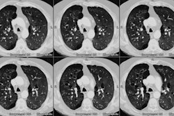
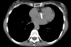
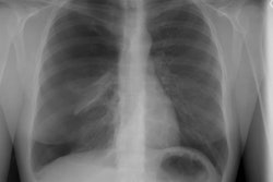
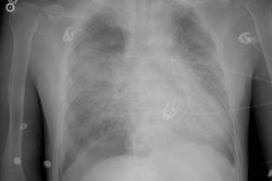
Cartilaginous disorders of the thorax can arise in the parenchyma, airways, chest wall, and axial skeleton. At radiography, pulmonary hamartoma is characterized by "popcorn" calcification or fat density, either of which is diagnostic. Bronchiectasis is best demonstrated at high-resolution computed tomography (CT) and has a "tramline" or "signet ring" appearance. Tracheopathia osteochondroplastica appears at CT as multiple sessile submucosal nodules with or without calcification along the cartilaginous portion of the trachea. In relapsing polychondritis, the trachea and mainstem bronchi have diffuse or focal thickening with luminal narrowing at radiography. Costochondritis of the chest wall has become more prevalent with increased intravenous drug abuse and may be demonstrated at CT as soft-tissue swelling along with underlying cartilaginous fragmentation and bone destruction. Enchondromas are expansile and may display a calcified cartilaginous matrix at radiography. In osteochondroma, the thickness of the cartilaginous cap determines the likelihood of malignant degeneration. At radiography, chondroblastomas have a round contour, sharp margins, and cortical scalloping, whereas chondrosarcomas are large masses with indistinct margins, cortical breakthrough, and soft-tissue extension. By identifying either a process affecting a cartilage-containing structure or a cartilaginous matrix within a lesion, the chest radiologist may be able to narrow the list of differential diagnostic possibilities substantially.
Publication Types:
Review
Review, tutorial
