J Nucl Med 2000 Jun;41(6):1010-5
FDG PET of recurrent or metastatic 131I-negative papillary
thyroid carcinoma.
Alnafisi NS, Driedger AA, Coates G, Moote DJ, Raphael SJ.
This study reports on the use of FDG PET in the follow-up of papillary thyroid
cancer patients with negative findings on 131I total body scans and elevated
levels of thyroglobulin after total thyroidectomy. METHODS: Eleven asymptomatic
patients with previous papillary thyroid cancer, total thyroidectomy, 131I
ablation, and treatment of all known metastases had negative findings on 131I
total body scans after therapy but persisting elevations of thyroglobulin when
not receiving thyroid hormone. All imaging before PET failed to show persisting
tumor. FDG PET was performed on all patients while receiving full thyroid
hormone replacement, except for the repeated scan of 1 patient (patient 6).
After the PET scan, all patients were referred for supplementary CT, sonography,
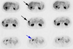
or biopsy of lesions in the neck. RESULTS: All 11 patients showed FDG uptake in
the neck or upper mediastinum-in the initial scan in 10 and in a repeated scan
in 1. Sonographically guided biopsy confirmed malignancy in 6, was nondiagnostic
in 2, and showed normal findings in 1. In 2 patients, the sonographic results
were normal and no biopsy was attempted. FDG imaging redirected the treatment of
7 patients, resulting in surgery and external beam radiotherapy in 3, surgery in
1, and external beam radiotherapy in 2. One patient declined further recommended
surgery. The other 4 patients remain under observation. Surgical histopathology
confirmed thyroid tumor in all 4 surgically treated patients. Retrospective
review of the original histopathology slides showed no preponderance of
aggressive histology. CONCLUSION: FDG PET is able to guide further evaluation of
thyroid cancer patients who have elevated thyroglobulin levels and normal
findings on 131I whole-body scanning.
