Radiology 2001 Jan;218(1):233-241
Complications (Excluding Hyperinflation) Involving the Native Lung after
Single-Lung Transplantation: Incidence, Radiologic Features, and Clinical Importance.
McAdams HP, Erasmus JJ, Palmer SM
PURPOSE: To determine the incidence, importance, and radiologic features of native lung
complications after single-lung transplantation. MATERIALS AND METHODS: Seventeen (15%) of
111 single-lung transplant recipients developed native lung complications (excluding
hyperinflation) 0-58 months (mean, 17 months) after transplantation. Complaints at
presentation, culture or histopathologic results, diagnostic or therapeutic procedures,
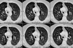
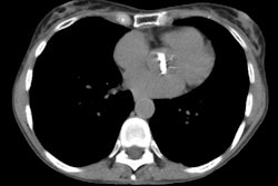
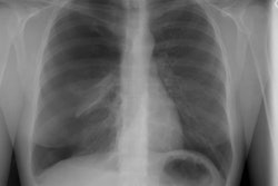
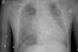
and outcome were recorded. Chest radiographs (n = 17) and computed tomographic (CT) scans
(n = 8) obtained at time of diagnosis were reviewed. Serial radiographs were assessed for
disease progression or improvement. RESULTS: The most common complications were infection
(n = 10), caused by bacteria (n = 4), fungi (n = 4), or mycobacteria (n = 2), typically
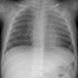
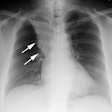
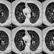
manifested as lobar or segmental opacities on chest radiographs or CT scans. Lung cancer
manifested as a solitary well-circumscribed nodule (n = 1), multiple nodules (n = 1), or a
hilar mass (n = 1). Five (29%) of 17 patients died of native lung complications. Seven
patients underwent mediastinoscopy (n = 3), lobectomy (n = 2), thoracoscopic wedge
resection (n = 2), tube thoracostomy (n = 2), or pneumonectomy (n = 1) for diagnosis or
treatment. CONCLUSION: Native lung complications occurred in 17 (15%) single-lung
transplant recipients, were most commonly due to infection or lung cancer, and caused
serious morbidity or mortality in 12 (71%) of 17 patients affected.
PMID: 11152808
