Diffuse Alveolar Damage:
Clinical:
Diffuse alveolar damage is a very common histologic pattern of lung injury (acute insult to the alveolar-capillary interface) and NOT a specific diagnosis [5]. DAD begins with damage to all three layers of the alveolar wall [5]. Endothelial damage permits leakage of plasma fluid into the interstitium and alveoli [5]. Epithelial and basement membrane damage is widespread with sloughing into the alveoli [5].It is found in a variety of disorders especially adult respiratory distress syndrome. Patients have a severe clinical course and typically require prolonged mechanical ventilation that can result in ventilator-induced lung injury [5].
Etiologies include: Infections (bacterial, viral, fungal), toxic inhalations, shock, drugs (bleomycin, busulfan, carmustine, cyclophosphamide), alveolar hemorrhage syndromes, acute hypersensitivity pneumonitis, and acute radiation reactions. In the absence of an identifiable cause DAD may be idiopathic - in which case the clinical diagnosis of acute interstitial pneumonia (Hamman-Rich syndrome) may be applied [4].
Acute respiratory deterioration due to DAD has also been described as an uncommon manifestation of connective tissue diseases including, polymyositis and dermatomyositis (most commonly and associated with a poor prognosis [3]), RA, SLE, and systemic sclerosis [3]. In patients with DAD and underlying ILD, the prognosis is worse with a mortality of between 75-86% [3,4].
Histologically diffuse alveolar damage is divided into two phases. In the early phases (from the initial injury through day 7) there is edema, epithelial necrosis and sloughing, a fibrinous exudate in the airspaces, and hyaline membrane formation [4] Hyaline membranes are homogeneous eosinophilic material composed of cellular debris, plasma proteins, and surfactant plastered against alveolar ducts and alveolar walls [4].
After the first week the organizing or repairative phase of DAD predominates - with mixed airspace and interstitial disease, alveolar type II pneumocyte hyperplasia and metaplasia and fibroblastic proliferation [4,5]. This can be followed by a chronic fibrotic phase if patients survive. The process is generally diffuse, but can be regional oand areas of sparing are common [4]. It is important to note that the two phases of DAD are not strictly sequential and there is a great deal of overlap between the two phases [4]. Alveolar collapse can be seen in both phases and accounts for striking volume loss in patients with DAD [4].
X-ray:
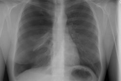
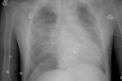
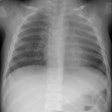
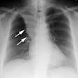
Chest radiographs performed during the early phase of DAD usually show a bilateral heterogeneous or homogeneous alveolar opacities often in a mid to lower lung zone distribution. Progression to diffuse opacification is common.
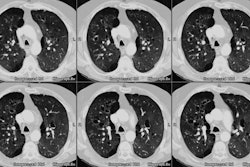
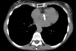
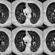
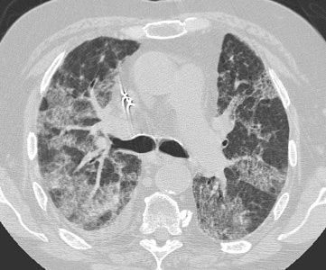
During the acute phase, CT of the chest will show rapidly progressive ground-glass opacities and/or consolidation bilaterally with septal thickening (producing a crazy-paving appearance) and areas of geographic sparing [4]. These findings reflect a combination of airspace exudates, interstitial edema, inflammation, and alveolar collapse that is usually most pronounced in the dependent portion of the lungs [4] and dependent atelectasis is common [5]. As the lung injury progresses, findings of fibrosis including reticulation, traction bronchiectasis, and honeycombing develop [4].
CT findings can return to normal in some patients, but between 38-85% have residual fibrosis that can be extensive and debilitating [4]. The fibrotic pattern is usually atypical involving less than 25% of the lung and it is most pronounced in the anterior nondependent portions of the lung (likely due to barotrauma from mechanical ventilation injury or oxygen toxicity- the dependent portions of the lung are protected by collapse) [4,5].Other findings include persistent ground-glass opacities, parenchymal bands, architectural distortion, and areas of permanent parenchymal destruction (pneumatoceles and focal emphysema) [5].
|
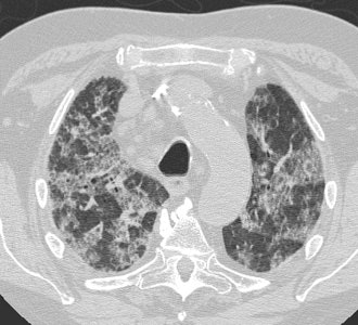
Diffuse Alveolar Damage: The patient below was admitted to the intensive care unit with increasing dyspnea. HRCT imaging revealed patchy areas of ground glass opacity and consolidation within the lungs bilaterally. There were prominent underlying reticulations within the areas of airspace disease. An open lung biopsy was performed and demonstrated diffuse alveolar damage. The etiology was not clinically apparent. |
  |
REFERENCES:
(1) J Thorac Imaging 1996; Anatomic distribution and histopathologic patterns in diffuse lung disease: correlation with HRCT. 11(1), 1-26 (No abstract available)
(2) Radiographics 2000; Rossi SE, et al. Pulmonary drug toxicity: Radiologic and pathologic manifestations. 20: 1245-1259
(3) Chest 2006; Parambil JG, et al. Diffuse alveolar damage. 130: 553-558
(4) Radiographics 2013; Kligerman SJ, et al. Organization and fibrosis as a response to lung injury in diffuse alveolar damage, organizing pneumonia, and acute fibrinous and organizing pneumonia. 33: 1951-1975
(5) Radiographics 2023; Marquis KM, et al. CT approach to lung injury. 43: e220176
