Correlation of chest radiographic findings with biopsy-proven acute lung rejection.
Kundu S, Herman SJ, Larhs A, Rappaport DC, Weisbrod GL, Maurer J, Chamberlain D, Winton T
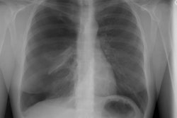
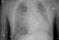
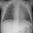
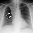
The purpose of this study was to determine the chest radiographic findings of acute rejection and the accuracy of chest radiography in making this diagnosis in patients undergoing lung transplantation. For each of 100 transbronchial biopsies performed on 25 lung transplant recipients (single lung in three, double lung in 22), chest radiographs obtained within 24 hours before the biopsy were reviewed retrospectively without knowledge of clinical or biopsy information. Transbronchial biopsy revealed 42 instances of acute rejection in 17 patients and 58 instances of no acute rejection (normal, n = 43; other processes, n = 15). All pulmonary parenchymal radiographic abnormalities were assessed. Acute rejection was associated with the presence of middle or lower lung reticular interstitial or airspace disease in 21 lungs (sensitivity = 0.50 [21/42]). This pattern was seen in 18 lungs without acute rejection (specificity = 0.69 [40/58]). There was no difference in the appearance of the lungs between grades 1 and 2 acute rejection. Normal lungs were noted in 20 instances of acute rejection (48%). The authors conclude that chest radiograph findings are abnormal in about 50% of instances of biopsy-proven acute rejection. Because the appearance of acute rejection is similar to that of other conditions, the diagnosis cannot be made accurately by chest radiography.
