Cancer 1998 May 1;82(9):1664-71
Primary staging and follow-up of high risk melanoma patients with whole-body
18F-fluorodeoxyglucose positron emission tomography: results of a prospective
study of 100 patients.
Rinne D, Baum RP, Hor G, Kaufmann R.
BACKGROUND: Positron emission tomography (PET) has been retrospectively reported
to be a sensitive method for detecting malignant melanoma metastases. METHODS:
One hundred consecutive patients with high risk melanoma (tumor thickness >
1.5 mm) were prospectively evaluated (52 at primary diagnosis, comprising Group
A, and 48 during follow-up, comprising Group B) by whole-body PET and
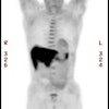
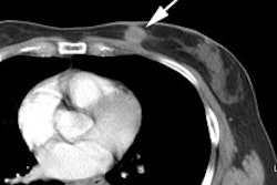
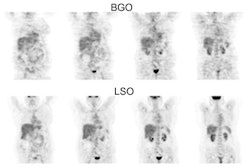
conventional diagnostics (CD). RESULTS: In Group A, the sensitivity of PET was
100% and the specificity was 94%, whereas CD did not identify any of the 9 lymph
node metastases and demonstrated a lower specificity (80%). In Group B, 121
lesions were detected, 111 by PET and 69 by conventional imaging. On the basis
of patients, the sensitivity, specificity, and accuracy of PET were 100%, 95.5%,
and 97.9%, respectively (91.8%, 94.4%, and 92.1%, respectively, on the basis of
single metastases). Prospectively, CD did not identify all patients with
progression (sensitivity, 84.6%) and detected significantly fewer metastases
(sensitivity, 57.5%) with much lower specificity (68.2% on the basis of
patients, 45% on the basis of single lesions); therefore, the accuracy of CD was
77.1% on the basis of patients and only 55.7% on the basis of single metastases.
Results also depended on specific sites: while PET yielded a higher sensitivity
in detecting cervical metastases (100% vs. 66.6%) and abdominal metastases (100%
vs. 26.6%), computed tomography proved to be superior in detecting small lung
metastases (87% vs. 69.6%). CONCLUSIONS: PET is a highly sensitive and specific
technique for melanoma staging. With the exception of the brain, one single
whole-body 18F-fluorodeoxyglucose-PET scan could replace the standard battery of
imaging tests currently performed on high risk melanoma patients.


