AJR Am J Roentgenol 1997 Sep;169(3):637-647. Radiology of obstructive pulmonary disease.
Webb WR
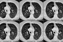
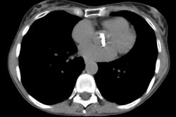
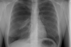
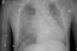
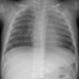
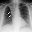
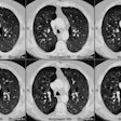
Obstructive lung diseases may be associated with a variety of pathologic findings, including emphysema, large airways abnormalities, and small airways abnormalities. The usefulness of plain radiographs for showing these findings is limited, although the presence of obstructive lung disease can often be inferred in the presence of increased lung volumes, gross lung destruction (emphysema) or large airways abnormalities. Furthermore, radiographs may not provide useful information in many patients with a known disease who experience an exacerbation of their symptoms. Nonetheless, chest radiographs are commonly obtained in this setting to assess the presence or absence of disease complications. HRCT can reveal morphologic abnormalities associated with obstructive lung disease with a greater accuracy than plain radiographs. HRCT is more sensitive than radiographs in showing emphysema, large airways abnormalities such as bronchiectasis, and small airways abnormalities such as bronchiolectasis and the tree-in-bud appearance. Furthermore, findings associated with abnormal ventilation, including mosaic perfusion, can also be seen on HRCT, as can findings of air trapping on expiratory scans. These latter findings are particularly helpful in diagnosing obstructive disease in the absence of other morphologic abnormalities. Although the indications for use of HRCT vary with the specific disease, HRCT can be valuable in patients in whom the diagnosis is, on the basis of clinical and plain radiographic findings, in question or for whom specific therapy is being contemplated.
PMID: 9275869, MUID: 97421752
