Staging of esophageal carcinoma:
T stage:
Tis- carcinoma in situ/high grade dysplasia
T1- carcinoma confined to the mucosa or submucosa
T1a invades lamina propria or muscularis mucosae
T1b invades submucosa
T2- tumor invades the muscularis propria
T3- tumor invades the adventitia of the esophageal wall
T4- tumor invades adjacent structures
T4a- potentially resectable tumors that invade adjacent structures such as the pleura-peritoneum, pericardium, or diaphragm
T4b- unresectable tumors that invade adjacent structures such as the aorta, carotid vessels, azygouos vein, trachea, left main bronchus, or vertebral body
N-stage:
N0- no regional lymph node mets
N1- 1-2 regional lymph node mets
N2- 3-6 regional lymph node mets
N3: More than 7 regional lymph node mets
M-stage:
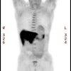
M0- Absent. Previously, non-regional lymph node metastases were considered M1a, such as mets to celiac nodes (lower thoracic tumor) or cervical nodes (upper esophageal primary tumor). M1a disease was associated with a worse prognosis. Patients with M1a disease who had a response to neoadjuvant therapy could still be candidates for esophagectomy and were potentially curable [2].
M1- Present
Overall staging is then further based on tumor histology
(squamous versus adenocarcinoma), tumor location (lower vs middle
and upper esophagus) and tumor grade (G1-G3). See the link below:
http://www.cancer.gov/cancertopics/pdq/treatment/esophageal/HealthProfessional/page3
REFERENCES:
(1) Radiol Clin N Am 2005; Korst RJ, Altorki NK. Imaging esophageal tumors. 43: 611-619
(2) Radiographics 2007; Bruzzi JF, et al. PET/CT of esophageal cancer: its role in clinical management. 27: 1635-1652
(3) Radiographics 2009; Kim TJ, et al. Multimodality
assessment of esophageal cancer: preoperative staging and
monitoring response to therapy. 29: 403-421
(4) Radiographics 2014; Hong SJ, et al. New TNM staging system for esophageal cancer: what chest radiologists need to know. 34: 1722-1740


